Explore
 Validate
 Learn
LearnMA5-18077
antibody from Invitrogen Antibodies
Targeting: SYNE1
8B, ARCA1, C6orf98, CPG2, dJ45H2.2, enaptin, KIAA0796, MYNE1, Nesp1, Nesprin-1, SCAR8, SYNE-1B
 Western blot
 Immunocytochemistry
Antibody data
- Antibody Data
- Antigen structure
- References [7]
- Comments [0]
- Validations
- Immunocytochemistry [3]
- Other assay [4]
Submit
Validation data
Reference
Comment
Report error
- Product number
- MA5-18077 - Provider product page

- Provider
- Invitrogen Antibodies
- Product name
- Nesprin 1 Monoclonal Antibody (MANNES1A(7A12))
- Antibody type
- Monoclonal
- Antigen
- Recombinant full-length protein
- Description
- This antibody recognizes the C-terminal region of nesprin-1. This target has a predicted molecular weight of 1011 kDa.
- Reactivity
- Human, Mouse, Rat, Rabbit
- Host
- Mouse
- Isotype
- IgG
- Antibody clone number
- MANNES1A(7A12)
- Vial size
- 100 μL
- Concentration
- 1 mg/mL
- Storage
- Store at 4°C short term. For long term storage, store at -20°C, avoiding freeze/thaw cycles.
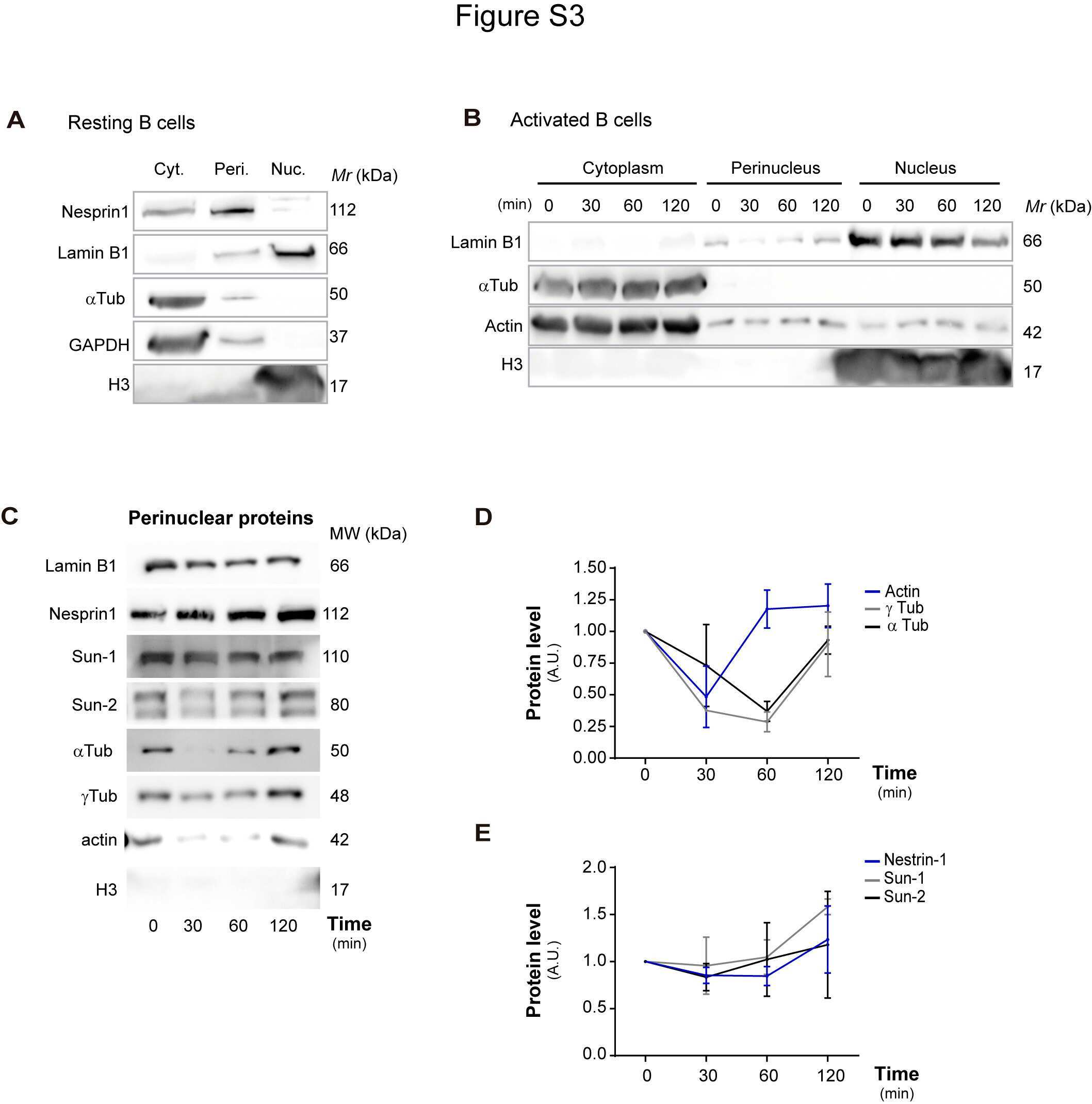
Submitted references B Cells Adapt Their Nuclear Morphology to Organize the Immune Synapse and Facilitate Antigen Extraction.
The LINC complex is required for endothelial cell adhesion and adaptation to shear stress and cyclic stretch.
Mechanical suppression of breast cancer cell invasion and paracrine signaling to osteoclasts requires nucleo-cytoskeletal connectivity.
Role of the nuclear membrane protein Emerin in front-rear polarity of the nucleus.
Nup93 regulates breast tumor growth by modulating cell proliferation and actin cytoskeleton remodeling.
Spatiotemporal Mislocalization of Nuclear Membrane-Associated Proteins in γ-Irradiation-Induced Senescent Cells.
Nuclear envelope rupture is induced by actin-based nucleus confinement.
Ulloa R, Corrales O, Cabrera-Reyes F, Jara-Wilde J, Saez JJ, Rivas C, Lagos J, Härtel S, Quiroga C, Yuseff MI, Diaz-Muñoz J
Frontiers in immunology 2021;12:801164
Frontiers in immunology 2021;12:801164
The LINC complex is required for endothelial cell adhesion and adaptation to shear stress and cyclic stretch.
Denis KB, Cabe JI, Danielsson BE, Tieu KV, Mayer CR, Conway DE
Molecular biology of the cell 2021 Aug 19;32(18):1654-1663
Molecular biology of the cell 2021 Aug 19;32(18):1654-1663
Mechanical suppression of breast cancer cell invasion and paracrine signaling to osteoclasts requires nucleo-cytoskeletal connectivity.
Yi X, Wright LE, Pagnotti GM, Uzer G, Powell KM, Wallace JM, Sankar U, Rubin CT, Mohammad K, Guise TA, Thompson WR
Bone research 2020 Nov 17;8(1):40
Bone research 2020 Nov 17;8(1):40
Role of the nuclear membrane protein Emerin in front-rear polarity of the nucleus.
Nastały P, Purushothaman D, Marchesi S, Poli A, Lendenmann T, Kidiyoor GR, Beznoussenko GV, Lavore S, Romano OM, Poulikakos D, Lagomarsino MC, Mironov AA, Ferrari A, Maiuri P
Nature communications 2020 May 1;11(1):2122
Nature communications 2020 May 1;11(1):2122
Nup93 regulates breast tumor growth by modulating cell proliferation and actin cytoskeleton remodeling.
Bersini S, Lytle NK, Schulte R, Huang L, Wahl GM, Hetzer MW
Life science alliance 2020 Jan;3(1)
Life science alliance 2020 Jan;3(1)
Spatiotemporal Mislocalization of Nuclear Membrane-Associated Proteins in γ-Irradiation-Induced Senescent Cells.
Svobodová Kovaříková A, Bártová E, Kovařík A, Lukášová E
Cells 2020 Apr 17;9(4)
Cells 2020 Apr 17;9(4)
Nuclear envelope rupture is induced by actin-based nucleus confinement.
Hatch EM, Hetzer MW
The Journal of cell biology 2016 Oct 10;215(1):27-36
The Journal of cell biology 2016 Oct 10;215(1):27-36
No comments: Submit comment
Supportive validation
- Submitted by
- Invitrogen Antibodies (provider)
- Main image

- Experimental details
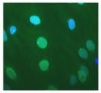
- Immunofluorescent analysis of Nesprin 1 in normal human fibroblast cells (green) using a Nesprin 1 monoclonal antibody (Product # MA5-18077); chromosomes were stained using DAPI (blue).
- Submitted by
- Invitrogen Antibodies (provider)
- Main image

- Experimental details
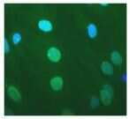
- Immunofluorescent analysis of Nesprin 1 in normal human fibroblast cells (green) using a Nesprin 1 monoclonal antibody (Product # MA5-18077); chromosomes were stained using DAPI (blue).
- Submitted by
- Invitrogen Antibodies (provider)
- Main image

- Experimental details
- Immunofluorescent analysis of Nesprin 1 in normal human fibroblast cells (green) using a Nesprin 1 monoclonal antibody (Product # MA5-18077); chromosomes were stained using DAPI (blue).

Supportive validation
- Submitted by
- Invitrogen Antibodies (provider)
- Main image
- Experimental details
- NULL
- Submitted by
- Invitrogen Antibodies (provider)
- Main image
- Experimental details
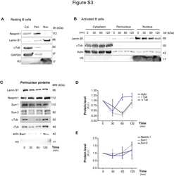
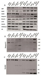
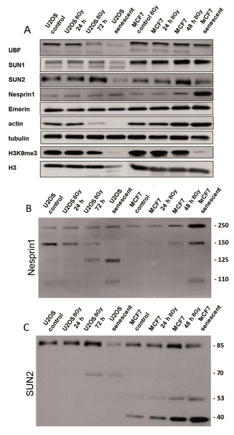
- Figure 6 Western blot analysis of protein expression levels ( A ) and their integrity ( B , C ) in control and irradiated MCF7 and U2OS cells. ( A ) Blot cuts showing signals of intact proteins. Note, reduction of SUN2 protein and near absence of H3K9me3 epigenetic modification in senescent cells (7 days post irradiation). ( B , C ) Reactivity of antibodies against nesprin-1 ( B ) and SUN2 ( C ) proteins on the whole blots. Note, significant fragmentation of intact nesprin-1 (250 kDa band) and SUN2 (85 kDa band) in irradiated cells.
- Submitted by
- Invitrogen Antibodies (provider)
- Main image
- Experimental details
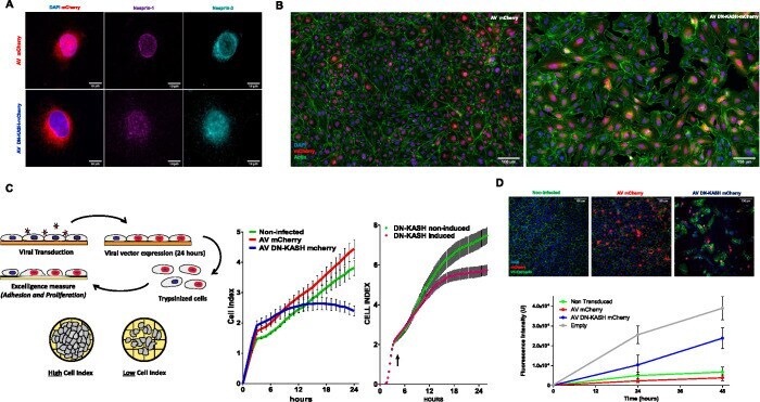
- FIGURE 1: DN-KASH expression disrupts the LINC complex and affects barrier formation. (A) Cells expressing adenovirus (AV) mCherry (red) or DN-KASH mCherry (red) were stained for nesprin-1 (magenta) and nesprin-2 (cyan). (B) Immunofluorescence of HUVEC-expressing mCherry adenovirus or DN-KASH-mCherry adenovirus after 48 h of growth. Cells were stained for DAPI (blue), actin (green), and mCherry (red). (C) Electrical impedance was measured for HUVEC infected by mCherry adenovirus (red) or DN-KASH-mCherry (blue) or noninfected control (green). HUVEC were seeded on Xcelligence plate (Roche) and impedance recorded over time (cell index). In a separate experiment, impedance was also measured for HUVEC infected by inducible DN-KASH-mCherry adenovirus. At 5 h, DN-KASH is induced with doxycycline (DN-KASH TetON) or left uninduced (DN-KASH TetOFF). Each condition is recorded for four different wells; error bars represent SE. Representative curves of three independent experiments. Representative images of three independent experiments. (D) Permissibility to Dextran-FITC 70 kDa was assessed for HUVEC infected by adenovirus-mCherry or DN-KASH-mCherry on 0.4 um Transwells. After 12 h, Dextran-FITC 70 kDa is added to the apical surface of cells. This point represents time 0 h. Basal fluorescence was measured 24 and 48 h later. After fixation, cells on Transwells were immunostained with DAPI and VE-cadherin antibody.
- Submitted by
- Invitrogen Antibodies (provider)
- Main image
- Experimental details
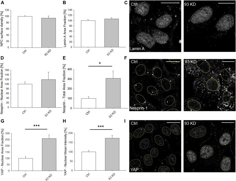
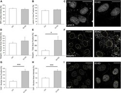
- Figure 2. NUP93 KD does not affect the structure of the nuclear envelope. (A, B) Quantification of nuclear pore complex surface density ( t test with P = 0.485, N >= 18 independent regions in three biological replicates, data normalized to ctrl cells) and (B) of lamin A area fraction ( t test with P = 0.335, N >= 13 independent regions in three biological replicates, data normalized to ctrl cells) after NUP93 KD. (C) Representative images of lamin A show no structural changes between ctrl and NUP93 KD BCCs. (D, F) Nesprin-1 analysis. (D, E) Quantification of nuclear (D) ( t test with P = 0.792, N >= 12 independent regions in three biological replicates, data normalized to ctrl cells) and total (E) ( t test with P < 0.05, N >= 4 independent regions in three biological replicates, data normalized to ctrl cells) Nesprin-1 area fraction after NUP93 KD. (F) Representative images showing that NUP93 KD increases Nesprin-1 expression without changes in terms of nuclear localization. Yellow lines are used to indicate cell nuclei. (G, H, I) YAP analysis. (G, H) Quantification of YAP area fraction (G) and mean fluorescence intensity (H) after NUP93 KD. t test with P < 0.001, N >= 14 independent regions in three biological replicates, data normalized to ctrl cells. (I) Representative images showing increased nuclear localization of YAP in NUP93 KD BCCs. Yellow lines are used to indicate cell nuclei. Scale bars: 10 mum. Source data are available for this figure. Source Data for Figure 2