Explore
 Validate
 Learn
Learn Immunohistochemistry
Antibody data
- Antibody Data
- Antigen structure
- References [0]
- Comments [0]
- Validations
- Immunohistochemistry [1]
- Flow cytometry [2]
Submit
Validation data
Reference
Comment
Report error
- Product number
- 10-4137-25 - Provider product page

- Provider
- ABEOMICS Inc.
- Product name
- Anti-CD28 Antibody
- Antibody type
- Monoclonal
- Description
- CD28 is a type I transmembrane protein that binds through its extracellular region to B7 proteins (CD80 and CD86), which are transmembrane proteins expressed on the surface of APCs (Antigen-Presenting Cells) and are up-regulated by inflammatory signals. CD28 mediates signals that promote T lymphocyte differentiation and proliferation, and enhance antibody production of B lymphocytes. Deficiencies in CD28 pathways result in complete T lymphocyte tolerance in vitro and in vivo. Both CD4+ and CD8+ memory T cells need CD28 costimulation to achieve maximal expansion and pathogen clearance. The blockade of the CD28-B7 interaction has been used to down-regulate the activation of the immune system in autoimmune diseases. Based on the expression of the costimulatory molecule CD28 on the surface of CD8+ T cells, two different lymphocyte subgroups have been designated: antigen-primed cytotoxic T cells (CD8+CD28+ T cells) and suppressor T cells (CD8+CD28â T cells). The frequency of CD28+CD8+T cells and especially the balance between CD8+CD28+ and CD8+CD28â T cells are important in many diseases, including CHB (Chronic Hepatitis B).
- Reactivity
- Human
- Host
- Mouse
- Conjugate
- Unconjugated
- Antigen sequence
A partial length recombinant protei
n from CD28 was used as the immunog
en for this antibody.- Isotype
- IgG
- Antibody clone number
- CB28
- Vial size
- 100 µg
- Concentration
- 0.5 mg/ml
- Storage
- Store the antibody at 4°C, stable for 6 months. For long-term storage, store at -20°C. Avoid repeat freez thawing
No comments: Submit comment
Supportive validation
- Submitted by
- ABEOMICS Inc. (provider)
- Main image
- Experimental details
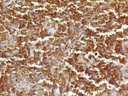
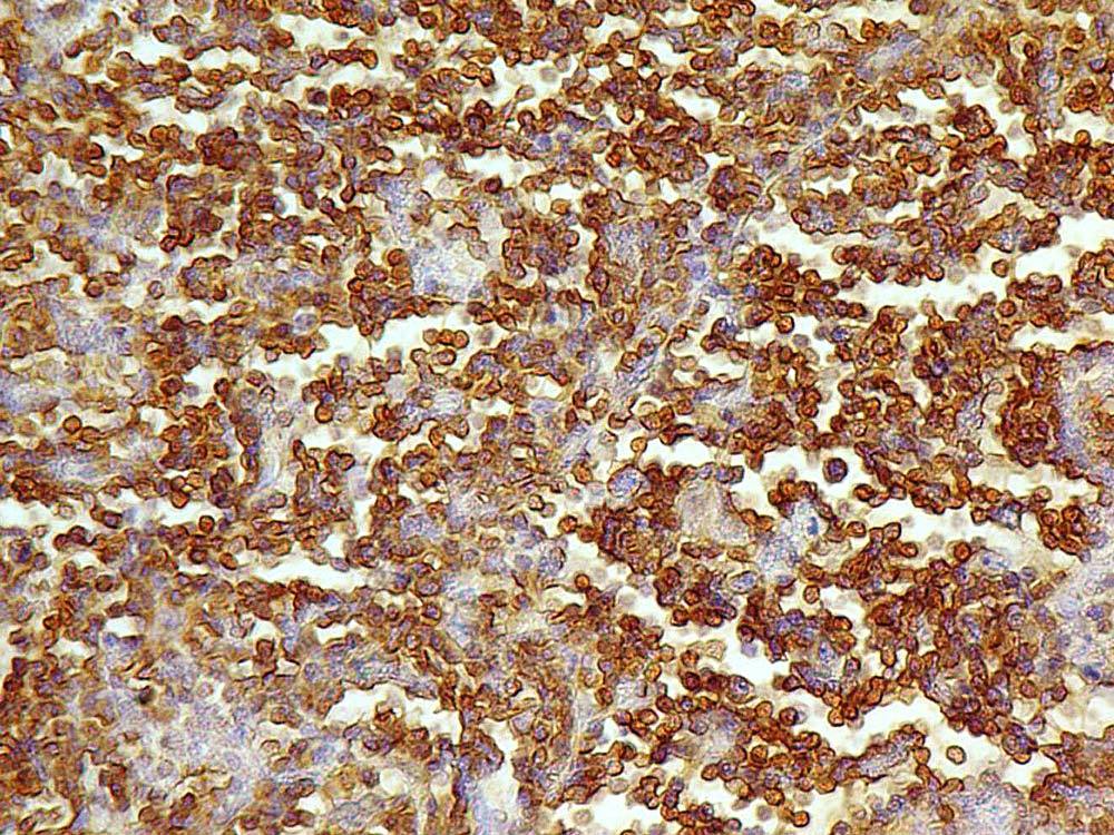
- Immunohistochemical analysis of CD 28 using Human Lungs Tissue using Anti-CD28 antibody (Clone:CB28) at 5 µg/ml.
- Protocol
- Protocol
Supportive validation
- Submitted by
- ABEOMICS Inc. (provider)
- Main image
- Experimental details
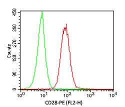
- Cell surface Flow analysis of hCD28 in Jurkat cells using 0.5 ug/ 10^6 cells. Green represents isotype control (ABEOMICS);red represents anti-hCD28 antibody (10-4137). Goat anti-mouse PE conjugated secondary antibody (ABEOMICS) was used. (Cells were incubated with primary antibody for 30 min. then washed twice with FLOW Staining buffer (ABEOMICS) by centrifuging at 1100 rpm for 5 min, followed by 30 min incubation with conjugated secondary antibody. Data acquisition was done after washing twice with Staining buffer).
- Protocol
- Protocol
- Submitted by
- ABEOMICS Inc. (provider)
- Main image

- Experimental details
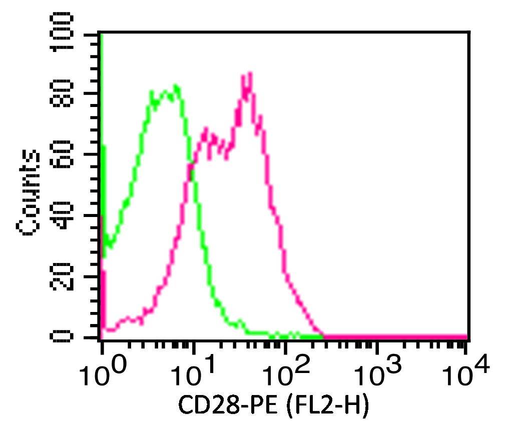
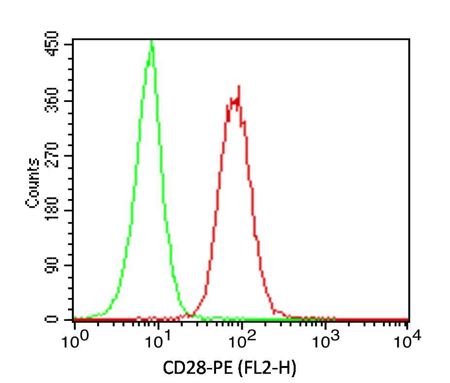
- Cell surface Flow analysis of CD28 in PBMC (lymphocytes gated) using 0.5 µg/ 10^6 cells. Green represents isotype control (ABEOMICS);red represents anti-hCD28 antibody (10-4137). Goat anti-mouse PE conjugated secondary antibody (ABEOMICS) was used. (Cells were incubated with primary antibody for 30 min. then washed twice with FLOW Staining buffer (ABEOMICS) by centrifuging at 1100 rpm for 5 min, followed by 30 min incubation with conjugated secondary antibody. Data acquisition was done after washing twice with Staining buffer).
- Protocol
- Protocol