Explore
 Validate
 Learn
LearnPA5-120369
antibody from Invitrogen Antibodies
Targeting: ARID1B
6A3-5, BAF250b, DAN15, ELD/OSA1, KIAA1235, p250R
 Western blot
 ELISA
ELISAAntibody data
- Antibody Data
- Antigen structure
- References [0]
- Comments [0]
- Validations
- Western blot [1]
- Immunocytochemistry [4]
- Immunohistochemistry [4]
Submit
Validation data
Reference
Comment
Report error
- Product number
- PA5-120369 - Provider product page

- Provider
- Invitrogen Antibodies
- Product name
- ARID1B Polyclonal Antibody
- Antibody type
- Polyclonal
- Antigen
- Recombinant protein fragment
- Description
- The target is usually found in the following locations: Nucleus.
- Reactivity
- Human, Mouse, Rat
- Host
- Rabbit
- Isotype
- IgG
- Vial size
- 100 μL
- Concentration
- 1.24 mg/mL
- Storage
- -20°C, Avoid Freeze/Thaw Cycles
No comments: Submit comment
Supportive validation
- Submitted by
- Invitrogen Antibodies (provider)
- Main image

- Experimental details
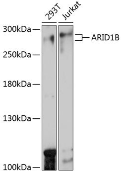
- Western blot analysis of ARID1B in extracts of various cell lines. Samples were incubated with ARID1B Polyclonal antibody (Product # PA5-120369) using a dilution of 1:1,000, followed by HRP Goat Anti-Rabbit IgG (H+L) at a dilution of 1:10,000. Lysates/proteins: 25 µg per lane. Blocking buffer: 3% nonfat dry milk in TBST. Detection: ECL Basic Kit. Exposure time: 30s.
Supportive validation
- Submitted by
- Invitrogen Antibodies (provider)
- Main image

- Experimental details
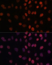
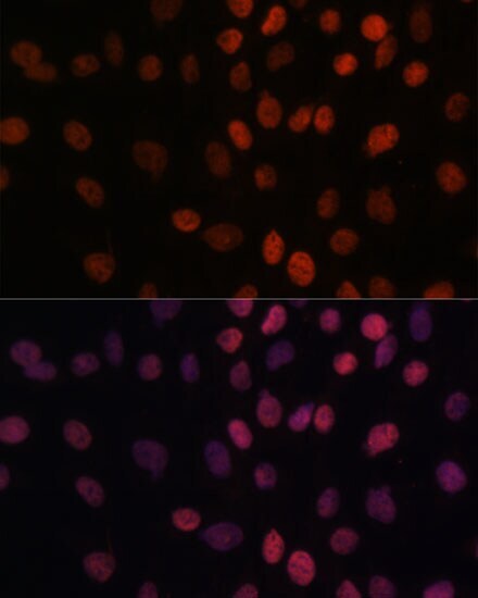
- Immunocytochemical analysis of ARID1B in C6 cells using a ARID1B Polyclonal antibody (Product # PA5-120369). Blue: DAPI for nuclear staining.

- Submitted by
- Invitrogen Antibodies (provider)
- Main image
- Experimental details
- Immunocytochemical analysis of ARID1B in C6 cells using a ARID1B Polyclonal antibody (Product # PA5-120369). Blue: DAPI for nuclear staining.

- Submitted by
- Invitrogen Antibodies (provider)
- Main image
- Experimental details
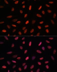
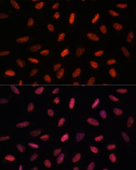
- Immunofluorescence analysis of ARID1B in C6 cells. Samples were incubated with ARID1B Polyclonal antibody (Product # PA5-120369) using a dilution of 1:100. Blue: DAPI for nuclear staining.
- Submitted by
- Invitrogen Antibodies (provider)
- Main image

- Experimental details
- Immunofluorescence analysis of ARID1B in U-2 OS cells. Samples were incubated with ARID1B Polyclonal antibody (Product # PA5-120369) using a dilution of 1:100. Blue: DAPI for nuclear staining.
Supportive validation
- Submitted by
- Invitrogen Antibodies (provider)
- Main image
- Experimental details
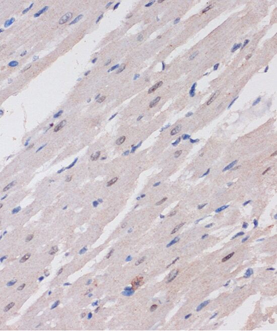
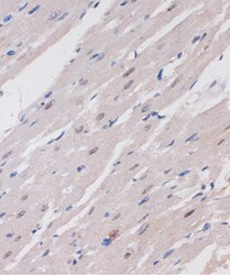
- Immunohistochemistry analysis of ARID1B in paraffin-embedded rat heart. Samples were incubated with ARID1B Polyclonal antibody (Product # PA5-120369) using a dilution of 1:100 (40x lens). Perform microwave antigen retrieval with 10 mM PBS buffer pH 7.2 before commencing with IHC staining protocol.
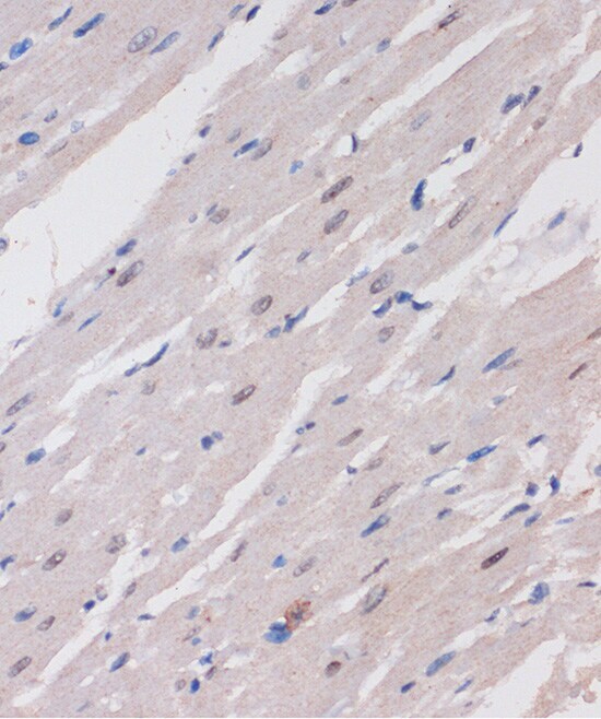
- Submitted by
- Invitrogen Antibodies (provider)
- Main image
- Experimental details
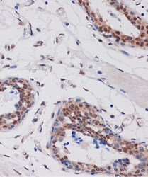
- Immunohistochemistry analysis of ARID1B in paraffin-embedded human breast. Samples were incubated with ARID1B Polyclonal antibody (Product # PA5-120369) using a dilution of 1:100 (40x lens). Perform microwave antigen retrieval with 10 mM PBS buffer pH 7.2 before commencing with IHC staining protocol.

- Submitted by
- Invitrogen Antibodies (provider)
- Main image
- Experimental details
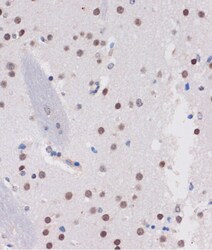
- Immunohistochemistry analysis of ARID1B in paraffin-embedded mouse brain. Samples were incubated with ARID1B Polyclonal antibody (Product # PA5-120369) using a dilution of 1:100 (40x lens). Perform microwave antigen retrieval with 10 mM PBS buffer pH 7.2 before commencing with IHC staining protocol.

- Submitted by
- Invitrogen Antibodies (provider)
- Main image
- Experimental details
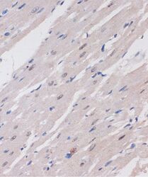
- Immunohistochemistry analysis of ARID1B in paraffin-embedded rat heart. Samples were incubated with ARID1B Polyclonal antibody (Product # PA5-120369) using a dilution of 1:100 (40x lens). Perform microwave antigen retrieval with 10 mM PBS buffer pH 7.2 before commencing with IHC staining protocol.