Explore
 Validate
 Learn
Learn Western blot
 Immunocytochemistry
Antibody data
- Antibody Data
- Antigen structure
- References [2]
- Comments [0]
- Validations
- Immunocytochemistry [1]
Submit
Validation data
Reference
Comment
Report error
- Product number
- HPA007599 - Provider product page

- Provider
- Atlas Antibodies
- Proper citation
- Atlas Antibodies Cat#HPA007599, RRID:AB_1854891
- Product name
- Anti-P4HA1
- Antibody type
- Polyclonal
- Description
- Polyclonal Antibody against Human P4HA1, Gene description: prolyl 4-hydroxylase, alpha polypeptide I, Alternative Gene Names: C-P4Halpha(I), P4HA, Validated applications: WB, ICC, IHC, Uniprot ID: P13674, Storage: Store at +4°C for short term storage. Long time storage is recommended at -20°C.
- Reactivity
- Human
- Host
- Rabbit
- Conjugate
- Unconjugated
- Isotype
- IgG
- Vial size
- 100 µl
- Concentration
- 0.2 mg/ml
- Storage
- Store at +4°C for short term storage. Long time storage is recommended at -20°C.
- Handling
- The antibody solution should be gently mixed before use.
Submitted references Lactate supports cell-autonomous ECM production to sustain metastatic behavior in prostate cancer
Prolyl 4-hydroxylase subunit alpha 1 (P4HA1) is a biomarker of poor prognosis in primary melanomas, and its depletion inhibits melanoma cell invasion and disrupts tumor blood vessel walls.
Ippolito L, Duatti A, Iozzo M, Comito G, Pardella E, Lorito N, Bacci M, Pranzini E, Santi A, Sandrini G, Catapano C, Serni S, Spatafora P, Morandi A, Giannoni E, Chiarugi P
EMBO Reports 2024;25(8):3506-3531
EMBO Reports 2024;25(8):3506-3531
Prolyl 4-hydroxylase subunit alpha 1 (P4HA1) is a biomarker of poor prognosis in primary melanomas, and its depletion inhibits melanoma cell invasion and disrupts tumor blood vessel walls.
Eriksson J, Le Joncour V, Jahkola T, Juteau S, Laakkonen P, Saksela O, Hölttä E
Molecular oncology 2020 Apr;14(4):742-762
Molecular oncology 2020 Apr;14(4):742-762
No comments: Submit comment
Supportive validation
- Submitted by
- Atlas Antibodies (provider)
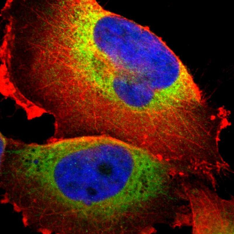
- Main image

- Experimental details
- Immunofluorescent staining of human cell line U-251 MG shows localization to endoplasmic reticulum.
- Sample type
- Human