Explore
 Validate
 Learn
Learn Western blot
 Immunocytochemistry
Antibody data
- Antibody Data
- Antigen structure
- References [1]
- Comments [0]
- Validations
- Immunocytochemistry [3]
- Immunohistochemistry [2]
- Other assay [1]
Submit
Validation data
Reference
Comment
Report error
- Product number
- PA5-21477 - Provider product page

- Provider
- Invitrogen Antibodies
- Product name
- BBOX1 Polyclonal Antibody
- Antibody type
- Polyclonal
- Antigen
- Recombinant full-length protein
- Description
- Recommended positive controls: Molt-4, mouse liver. Predicted reactivity: Mouse (86%), Rat (85%), Bovine (84%). Store product as a concentrated solution. Centrifuge briefly prior to opening the vial.
- Reactivity
- Human, Mouse
- Host
- Rabbit
- Isotype
- IgG
- Vial size
- 100 μL
- Concentration
- 0.76 mg/mL
- Storage
- Store at 4°C short term. For long term storage, store at -20°C, avoiding freeze/thaw cycles.
Submitted references Spatiotemporal gene expression patterns reveal molecular relatedness between retinal laminae.
Jiang D, Burger CA, Casasent AK, Albrecht NE, Li F, Samuel MA
The Journal of comparative neurology 2020 Apr 1;528(5):729-755
The Journal of comparative neurology 2020 Apr 1;528(5):729-755
No comments: Submit comment
Supportive validation
- Submitted by
- Invitrogen Antibodies (provider)
- Main image
- Experimental details
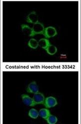
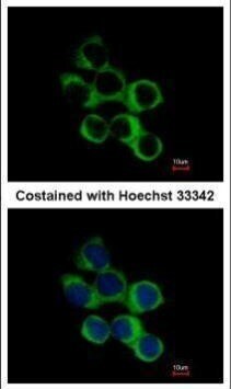
- Immunofluorescent analysis of Gamma-butyrobetaine dioxygenase in methanol-fixed A431 cells using a Gamma-butyrobetaine dioxygenase polyclonal antibody (Product # PA5-21477) at a 1:500 dilution.
- Submitted by
- Invitrogen Antibodies (provider)
- Main image
- Experimental details
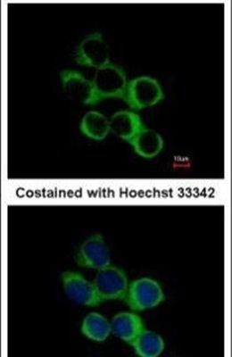
- Immunofluorescence analysis of methanol-fixed A431, using BBOX1 antibody (Product # PA5-21477) at 1:500 dilution.
- Submitted by
- Invitrogen Antibodies (provider)
- Main image

- Experimental details
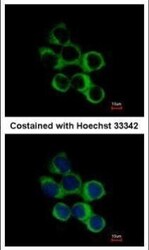
- Immunofluorescence analysis of methanol-fixed A431, using BBOX1 antibody (Product # PA5-21477) at 1:500 dilution.

Supportive validation
- Submitted by
- Invitrogen Antibodies (provider)
- Main image
- Experimental details
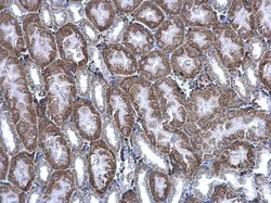
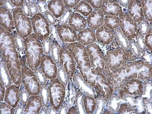
- BBOX1 Polyclonal Antibody detects BBOX1 protein at cytoplasm on mouse kidney by immunohistochemical analysis. Sample: Paraffin-embedded mouse kidney. BBOX1 Polyclonal Antibody (Product # PA5-21477) diluted at 1:500. Antigen Retrieval: EDTA based buffer, pH 8.0, 15 min.
- Submitted by
- Invitrogen Antibodies (provider)
- Main image
- Experimental details
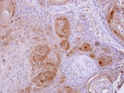
- BBOX1 Polyclonal Antibody detects BBOX1 protein at cytosol on Ca922 xenograft by immunohistochemical analysis. Sample: Paraffin-embedded Ca922 xenograft. BBOX1 Polyclonal Antibody (Product # PA5-21477) dilution: 1:500. Antigen Retrieval: EDTA based buffer, pH 8.0, 15 min.

Supportive validation
- Submitted by
- Invitrogen Antibodies (provider)
- Main image
- Experimental details
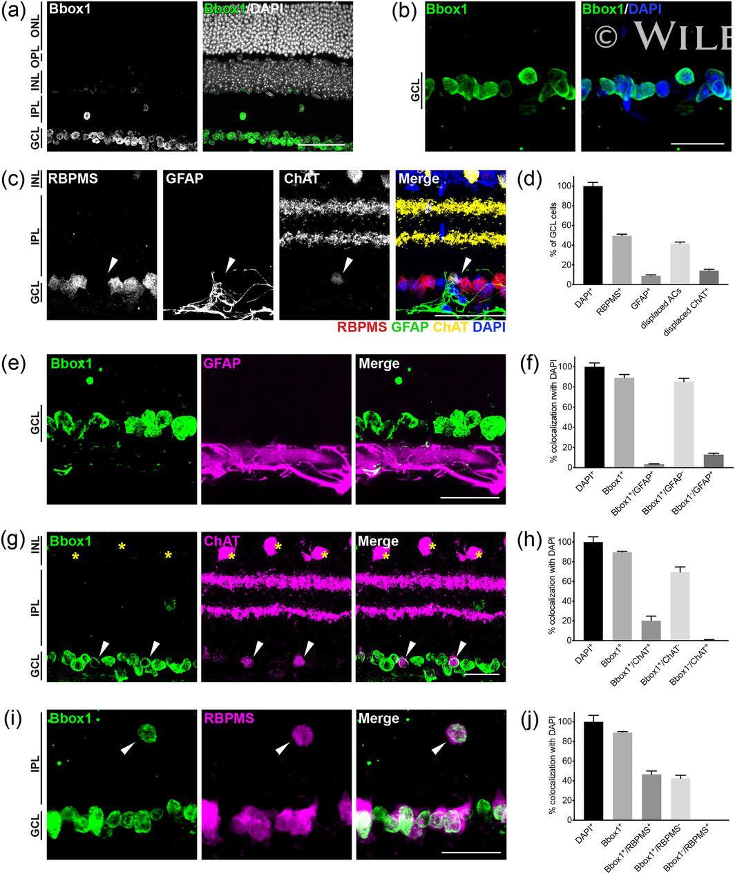
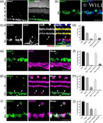
- Bbox1 labels the ganglion cell layer (GCL) in adult retina. (a,b) Immunohistochemical images of Bbox1 (green) at lower (a) and higher (b) magnification in 14-week-old retina reveal that Bbox1 is exclusively localized to the GCL. ONL, outer nuclear layer; OPL, outer plexiform layer; INL, inner nuclear layer; IPL, inner plexiform layer. (c) Representative images showing the major cell classes residing in the adult GCL: RGCs (RBPMS), astrocytes (GFAP), and displaced starburst amacrines (ChAT; white arrows). (d) Quantification of the major cell classes in the GCL in adult 14-week-old retina (RBPMS, retinal ganglion cells; GFAP, astrocytes; ChAT, displaced amacrine cells). n = 3 animals. (e,f) Representative images (e) and quantification (f) of Bbox1 (green) and astrocytes (magenta) colocalization in adult animals. Bbox1 shows very little overlap with astrocytes, which reside below GCL neurons. (g,h) Representative images (g) and quantification (h) of Bbox1 (green) and ChAT amacrine costaining in adult retina. Bbox1 does not label ChAT amacrines in the INL (yellow asterisks) but does label displaced ChAT amacrines in the GCL (white arrows). (i,j) Representative images (i) and quantification (j) of Bbox1 (green) and ganglion cells (RBPMS, magenta) colocalization in adult retina. Bbox1 labels all ganglion cells in the GCL and also labels displaced ganglion cells in the INL (white arrows). n = 3 animals. Data are represented as the mean +- SEM . Scale bars = 50 mum [Color figure can