Explore
 Validate
 Learn
Learn Western blot
Antibody data
- Antibody Data
- Antigen structure
- References [0]
- Comments [0]
- Validations
- Western blot [3]
- Immunocytochemistry [2]
- Immunohistochemistry [4]
Submit
Validation data
Reference
Comment
Report error
- Product number
- GTX115969 - Provider product page

- Provider
- GeneTex
- Proper citation
- GeneTex Cat#GTX115969, RRID:AB_10620389
- Product name
- QKI antibody [N2C3]
- Antibody type
- Polyclonal
- Reactivity
- Human, Mouse, Rat
- Host
- Rabbit
No comments: Submit comment
Supportive validation
- Submitted by
- GeneTex (provider)
- Main image
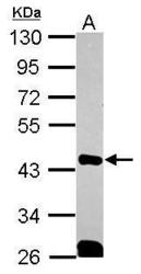
- Experimental details
- Sample (30 ug of whole cell lysate) A: 293T 10% SDS PAGE GTX115969 diluted at 1:1000

- Submitted by
- GeneTex (provider)
- Main image
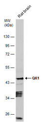
- Experimental details
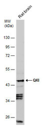
- Rat tissue extract (50 ?g) was separated by 10% SDS-PAGE, and the membrane was blotted with QKI antibody [N2C3] (GTX115969) diluted at 1:1000.
- Submitted by
- GeneTex (provider)
- Main image
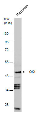
- Experimental details
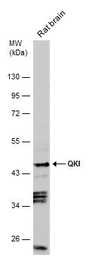
- Rat tissue extract (50 ?g) was separated by 10% SDS-PAGE, and the membrane was blotted with QKI antibody [N2C3] (GTX115969) diluted at 1:1000.
Supportive validation
- Submitted by
- GeneTex (provider)
- Main image
- Experimental details
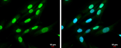
- Immunofluorescence analysis of paraformaldehyde-fixed A431, using QK1(GTX115969) antibody at 1:500 dilution.

- Submitted by
- GeneTex (provider)
- Main image
- Experimental details
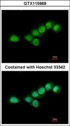
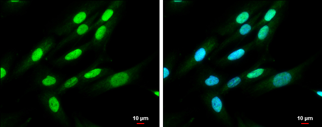
- QKI antibody [N2C3] detects QKI protein at cytoplasm and nucleus by immunofluorescent analysis.Sample: SK-N-SH cells were fixed in 4% paraformaldehyde at RT for 15 min.Green: QKI protein stained by QKI antibody [N2C3] (GTX115969) diluted at 1:500.Blue: Hoechst 33342 staining.Scale bar = 10 £gm.
Supportive validation
- Submitted by
- GeneTex (provider)
- Main image
- Experimental details
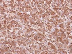
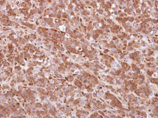
- Immunohistochemical analysis of paraffin-embedded MDA-MB157 xenograft, using QKI (GTX115969) antibody at 1:100 dilution.
- Submitted by
- GeneTex (provider)
- Main image
- Experimental details
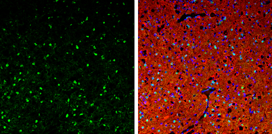
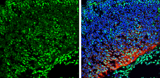
- QKI antibody [N2C3] detects QKI protein expression by immunohistochemical analysis.Sample: Frozen sectioned E13.5 Rat brain. Green: QKI protein stained by QKI antibody [N2C3] (GTX115969) diluted at 1:250.Red: beta Tubulin 3/ TUJ1, a mature neuron marker, stained by beta Tubulin 3/ TUJ1 antibody [GT11710] (GTX631836) diluted at 1:500.Blue: Fluoroshield with DAPI (GTX30920).
- Submitted by
- GeneTex (provider)
- Main image

- Experimental details
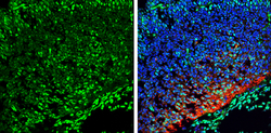
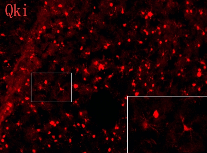
- QKI antibody [N2C3] detects QKI proteins on embryonic mouse brain by immunohistochemical analysis. Sample: Frozen section of embryonic mouse brain (mE18.5) Red:QKI antibody [N2C3] (GTX115969) diluted at 1:200.
- Submitted by
- GeneTex (provider)
- Main image

- Experimental details
- QKI antibody [N2C3] detects QKI protein expression by immunohistochemical analysis.Sample: Frozen-sectioned adult mouse cerebellum. Green: QKI protein stained by QKI antibody [N2C3] (GTX115969) diluted at 1:250.Red: beta Tubulin 3/ TUJ1, stained by beta Tubulin 3/ TUJ1 antibody [GT11710] (GTX631836) diluted at 1:500.Blue: Fluoroshield with DAPI (GTX30920).