 Explore
 Validate
 Learn
Learn Western blot
 Immunocytochemistry
Antibody data
- Antibody Data
- Antigen structure
- References [1]
- Comments [0]
- Validations
- Immunocytochemistry [2]
- Immunohistochemistry [3]
- Other assay [2]
Submit
Validation data
Reference
Comment
Report error
- Product number
- PA5-30563 - Provider product page

- Provider
- Invitrogen Antibodies
- Product name
- QKI Polyclonal Antibody
- Antibody type
- Polyclonal
- Antigen
- Recombinant full-length protein
- Description
- Recommended positive controls: 293T, rat brain. Predicted reactivity: Xenopus laevis (93%), Dog (100%), Cat (100%), Pig (100%), Chicken (98%), Bovine (100%). Store product as a concentrated solution. Centrifuge briefly prior to opening the vial.
- Reactivity
- Human, Mouse, Rat
- Host
- Rabbit
- Isotype
- IgG
- Vial size
- 100 μL
- Concentration
- 0.99 mg/mL
- Storage
- Store at 4°C short term. For long term storage, store at -20°C, avoiding freeze/thaw cycles.
Submitted references Quantitative proteomics identifies altered O-GlcNAcylation of structural, synaptic and memory-associated proteins in Alzheimer's disease.
Wang S, Yang F, Petyuk VA, Shukla AK, Monroe ME, Gritsenko MA, Rodland KD, Smith RD, Qian WJ, Gong CX, Liu T
The Journal of pathology 2017 Sep;243(1):78-88
The Journal of pathology 2017 Sep;243(1):78-88
No comments: Submit comment
Supportive validation
- Submitted by
- Invitrogen Antibodies (provider)
- Main image
- Experimental details
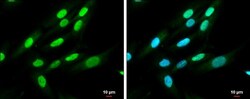
- Immunocytochemistry-Immunofluorescence analysis of QKI was performed in SK-N-SH cells fixed in 4% paraformaldehyde at RT for 15 min. Green: QKI Polyclonal Antibody (Product # PA5-30563) diluted at 1:500. Blue: Hoechst 33342 staining. Scale bar = 10 µm.

- Submitted by
- Invitrogen Antibodies (provider)
- Main image

- Experimental details
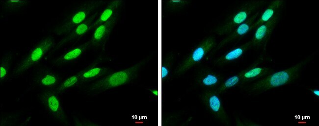
- Immunocytochemistry-Immunofluorescence analysis of QKI was performed in SK-N-SH cells fixed in 4% paraformaldehyde at RT for 15 min. Green: QKI Polyclonal Antibody (Product # PA5-30563) diluted at 1:500. Blue: Hoechst 33342 staining. Scale bar = 10 µm.
Supportive validation
- Submitted by
- Invitrogen Antibodies (provider)
- Main image
- Experimental details
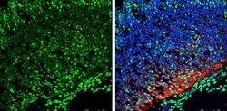
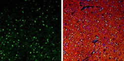
- Immunohistochemistry (Frozen) analysis of QKI was performed in frozen sectioned E13.5 Rat brain tissue using QKI Polyclonal Antibody (Product # PA5-30563) at a dilution of 1:250 (Green). Red: beta Tubulin 3/ TUJ1, a mature neuron marker, stained by beta Tubulin 3/ TUJ1 antibody diluted at 1:500. Blue: Fluoroshield with DAPI.
- Submitted by
- Invitrogen Antibodies (provider)
- Main image
- Experimental details
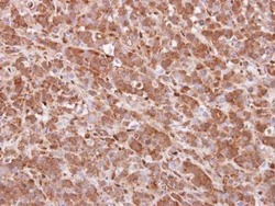
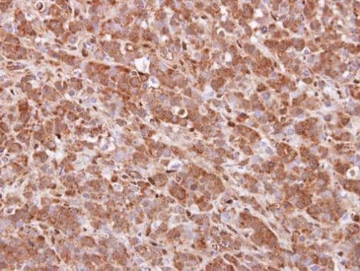
- Immunohistochemical analysis of paraffin-embedded MDA-MB157 xenograft, using QKI (Product # PA5-30563) antibody at 1:100 dilution. Antigen Retrieval: EDTA based buffer, pH 8.0, 15 min.
- Submitted by
- Invitrogen Antibodies (provider)
- Main image
- Experimental details
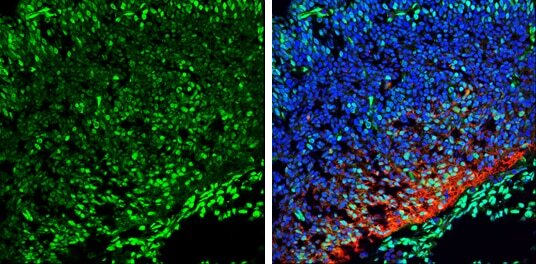
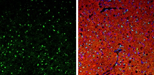
- Immunohistochemistry (Frozen) analysis of QKI was performed in frozen-sectioned adult mouse cerebellum tissue using QKI Polyclonal Antibody (Product # PA5-30563) at a dilution of 1:250 (Green). Red: beta Tubulin 3/ TUJ1, stained by beta Tubulin 3/ TUJ1 antibody diluted at 1:500. Blue: Fluoroshield with DAPI.
Supportive validation
- Submitted by
- Invitrogen Antibodies (provider)
- Main image
- Experimental details
- NULL

- Submitted by
- Invitrogen Antibodies (provider)
- Main image
- Experimental details
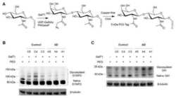
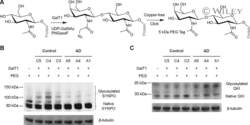
- Verification of O-GlcNAc quantification with western blotting analysis using lysate of the human brain tissue. (A) PEG labelling of O-GlcNAcylated proteins. First, the O-GlcNAc motif was labelled with GalNAz by GalT1 and UDP-GalNAz, and the N-glycans were removed by PNGase F. Second, the azide group was linked to a 5-kDa PEG tag through copper-free click chemistry. (B and C) The proteins were precipitated and separated by SDS-PAGE, and detected with antibody against SYNPO (B) or antibody against protein quaking (QKI) (C) through western blotting. beta-Tubulin was used as a loading control.
