 Explore
 Validate
 Learn
Learn39-7300
antibody from Invitrogen Antibodies
Targeting: RIOX2
FLJ14393, JMJD10, mdig, MINA, MINA53, NO52
 Western blot
 ELISA
ELISA Immunocytochemistry
Antibody data
- Antibody Data
- Antigen structure
- References [7]
- Comments [0]
- Validations
- Immunocytochemistry [2]
- Other assay [4]
Submit
Validation data
Reference
Comment
Report error
- Product number
- 39-7300 - Provider product page

- Provider
- Invitrogen Antibodies
- Product name
- MINA53 Monoclonal Antibody (M532)
- Antibody type
- Monoclonal
- Antigen
- Recombinant full-length protein
- Reactivity
- Human
- Host
- Mouse
- Isotype
- IgG
- Antibody clone number
- M532
- Vial size
- 100 μg
- Concentration
- 0.5 mg/mL
- Storage
- -20°C
Submitted references Environmentally-induced mdig contributes to the severity of COVID-19 through fostering expression of SARS-CoV-2 receptor NRPs and glycan metabolism.
ZNF143-Mediated H3K9 Trimethylation Upregulates CDC6 by Activating MDIG in Hepatocellular Carcinoma.
CRISPR-Cas9 gene editing causes alternative splicing of the targeting mRNA.
MINA53 deficiency leads to glioblastoma cell apoptosis via inducing DNA replication stress and diminishing DNA damage response.
Transcriptional activation of Mina by Sp1/3 factors.
Mina, an Il4 repressor, controls T helper type 2 bias.
Increased expression of a Myc target gene Mina53 in human colon cancer.
Zhang Q, Wadgaonkar P, Xu L, Thakur C, Fu Y, Bi Z, Qiu Y, Almutairy B, Zhang W, Stemmer P, Chen F
Theranostics 2021;11(16):7970-7983
Theranostics 2021;11(16):7970-7983
ZNF143-Mediated H3K9 Trimethylation Upregulates CDC6 by Activating MDIG in Hepatocellular Carcinoma.
Zhang L, Huo Q, Ge C, Zhao F, Zhou Q, Chen X, Tian H, Chen T, Xie H, Cui Y, Yao M, Li H, Li J
Cancer research 2020 Jun 15;80(12):2599-2611
Cancer research 2020 Jun 15;80(12):2599-2611
CRISPR-Cas9 gene editing causes alternative splicing of the targeting mRNA.
Zhang Q, Fu Y, Thakur C, Bi Z, Wadgaonkar P, Qiu Y, Xu L, Rice M, Zhang W, Almutairy B, Chen F
Biochemical and biophysical research communications 2020 Jul 12;528(1):54-61
Biochemical and biophysical research communications 2020 Jul 12;528(1):54-61
MINA53 deficiency leads to glioblastoma cell apoptosis via inducing DNA replication stress and diminishing DNA damage response.
Xuan F, Huang M, Zhao E, Cui H
Cell death & disease 2018 Oct 17;9(11):1062
Cell death & disease 2018 Oct 17;9(11):1062
Transcriptional activation of Mina by Sp1/3 factors.
Lian S, Potula HH, Pillai MR, Van Stry M, Koyanagi M, Chung L, Watanabe M, Bix M
PloS one 2013;8(12):e80638
PloS one 2013;8(12):e80638
Mina, an Il4 repressor, controls T helper type 2 bias.
Okamoto M, Van Stry M, Chung L, Koyanagi M, Sun X, Suzuki Y, Ohara O, Kitamura H, Hijikata A, Kubo M, Bix M
Nature immunology 2009 Aug;10(8):872-9
Nature immunology 2009 Aug;10(8):872-9
Increased expression of a Myc target gene Mina53 in human colon cancer.
Teye K, Tsuneoka M, Arima N, Koda Y, Nakamura Y, Ueta Y, Shirouzu K, Kimura H
The American journal of pathology 2004 Jan;164(1):205-16
The American journal of pathology 2004 Jan;164(1):205-16
No comments: Submit comment
Supportive validation
- Submitted by
- Invitrogen Antibodies (provider)
- Main image
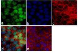
- Experimental details
- Immunofluorescence analysis of MINA53 was performed using 70% confluent log phase HCT 116 cells. The cells were fixed with 4% paraformaldehyde for 10 minutes, permeabilized with 0.1% Triton X-100 for 10 minutes, and blocked with 1% BSA for 1 hour at room temperature. The cells were labeled with MINA53 (M532) Mouse Monoclonal Antibody (Product # 39-7300) at 2 µg/mL in 0.1% BSA and incubated for 3 hours at room temperature and then labeled with Goat anti-Mouse IgG (H+L) Superclonal Secondary Antibody, Alexa Fluor® 488 conjugate (Product # A28175) at a dilution of 1:2000 for 45 minutes at room temperature (Panel a: green). Nuclei (Panel b: blue) were stained with SlowFade® Gold Antifade Mountant with DAPI (Product # S36938). F-actin (Panel c: red) was stained with Rhodamine Phalloidin (Product # R415, 1:300). Panel d represents the merged image showing nuclear localization. Panel e shows the no primary antibody control. The images were captured at 60X magnification.
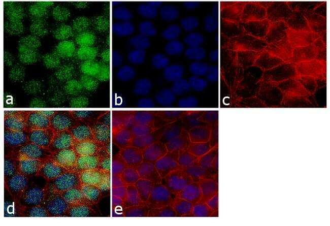
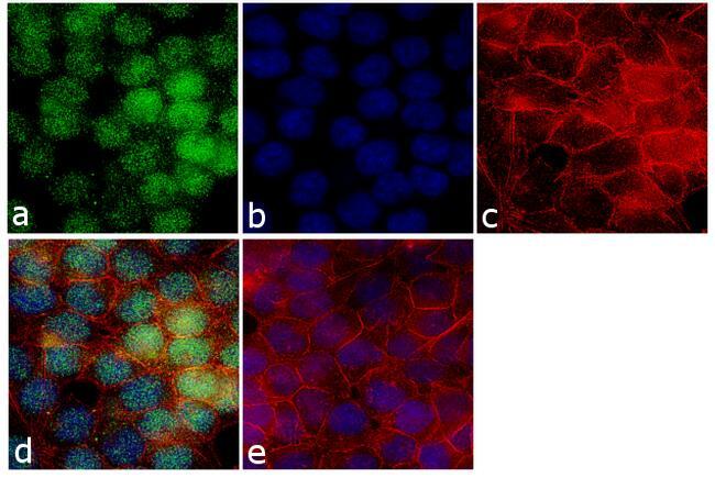
Supportive validation
- Submitted by
- Invitrogen Antibodies (provider)
- Main image
- Experimental details
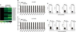
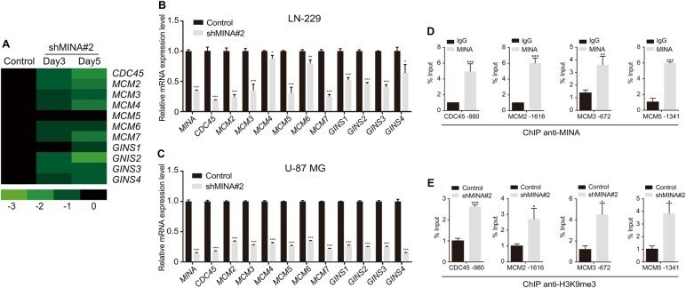
- Fig. 2 MINA53 regulates the CMG complex genes expression. a Expression alteration of CMG genes in LN-229 cells after MINA53 knockdown for 3 or 5 days. b mRNA expression of the indicated genes in LN-229 and c U-87 MG cells with or without MINA53 knockdown. d ChIP-qPCR analysis of MINA53 enrichment at the promoter regions of CDC45 , MCM2 , MCM3 , or MCM5 in LN-229 cells. e ChIP-qPCR analysis of H3K9me3 enrichment at the promoter regions of CDC45 , MCM2 , MCM3 , or MCM5 in LN-229 cells with or without MINA53 knockdown. All data were analyzed using two-tailed Student's t -tests. Mean +- s.e.m., n = 3, * P < 0.05, ** P < 0.01, *** P < 0.001
- Submitted by
- Invitrogen Antibodies (provider)
- Main image
- Experimental details
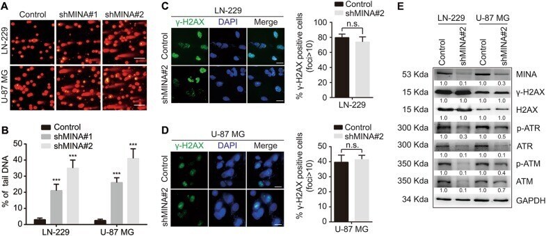
- Fig. 3 MINA53 deficiency induces DNA damage and reduces the activity of the ATM/ATR-H2AX pathway. a Representative fluorescent micrographs of comet assays and b quantification of the percentage of tail DNA of the indicated cells. Scale bar= 15 µm. c, d Immunofluorescence staining of γ-H2AX and quantification of γ-H2AX-positive cells of the indicated cells. Scale bar= 5 µm. e Western blot analysis of the indicated proteins in LN-229 and U-87 MG cells with or without MINA53 knockdown. All data were analyzed using two-tailed Student’s t-tests. Mean s.e.m., n= 3, ***P< 0.001
- Submitted by
- Invitrogen Antibodies (provider)
- Main image
- Experimental details
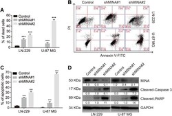
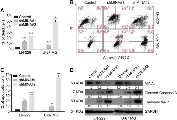
- Fig. 4 Knockdown of MINA53 induces glioblastoma cell apoptosis. a Quantification of the percentage of dead cells of the indicated cells. b Flow cytometry assays analyzing cell apoptosis and c quantification of the percentage of apoptosis cells of LN-229 and U-87 MG cells with or without MINA53 knockdown. d Western blot analysis of the indicated proteins in LN-229 and U-87 MG cells with or without MINA53 knockdown. All data were analyzed using two-tailed Student's t -tests. Mean +- s.e.m., n = 3, *** P < 0.001, n.s. no significance
- Submitted by
- Invitrogen Antibodies (provider)
- Main image
- Experimental details
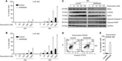
- Fig. 5 MINA53 deficiency sensitizes glioblastoma cells to Doxorubicin. U-87 MG cells with or without MINA53 knockdown were stimulated with Doxorubicin at indicated concentrations for 24 or 48 h. a Quantification of the percentage of dead cells and b the percentage of tail DNA in the indicated cells. c Western blot analysis of the indicated proteins after U-87 MG cells with or without MINA53 knockdown were stimulated with Doxorubicin at the indicated concentrations for 48 h. d Flow cytometry assays analyzing cell apoptosis and e quantification of the percentage of apoptosis cells of U-87 MG cells with or without MINA53 knockdown after stimulated with 500 nM Doxorubicin for 48 h. All data were analyzed using two-tailed Student's t -tests. Mean +- s.e.m., n = 3, *** P < 0.001
