 Explore
 Validate
 Learn
Learn Western blot
 Immunocytochemistry
Antibody data
- Antibody Data
- Antigen structure
- References [8]
- Comments [0]
- Validations
- Immunocytochemistry [4]
- Other assay [3]
Submit
Validation data
Reference
Comment
Report error
- Product number
- PA1-1962 - Provider product page

- Provider
- Invitrogen Antibodies
- Product name
- PSMA5 Polyclonal Antibody
- Antibody type
- Polyclonal
- Antigen
- Synthetic peptide
- Description
- PA1-1962 detects proteasome 20S subunit alpha 5 from human, rat, mouse and hamster cell lysates. PA1-1962 has been successfully used in Western blot and immunofluorescence procedures. By Western blot, this antibody detects a 26.5 kDa protein representing proteasome 20S subunit alpha 5 from human, rat, mouse, and hamster cell extracts. PA1-1962 immunogen is a synthetic peptide corresponding to residues C A(120) L Q F G E E D A D P G A M(133) of human proteasome 20S subunit alpha 5. PA1-1962 immunizing peptide (Cat. # PEP-210) is available for use in neutralization and control experiments.
- Reactivity
- Human, Mouse, Rat, Hamster
- Host
- Rabbit
- Isotype
- IgG
- Vial size
- 100 μg
- Concentration
- 1 mg/mL
- Storage
- -20°C, Avoid Freeze/Thaw Cycles
Submitted references Active Protein Neddylation or Ubiquitylation Is Dispensable for Stress Granule Dynamics.
Network Analysis of UBE3A/E6AP-Associated Proteins Provides Connections to Several Distinct Cellular Processes.
The transition zone protein Rpgrip1l regulates proteasomal activity at the primary cilium.
ATM regulates proteasome-dependent subnuclear localization of TRF1, which is important for telomere maintenance.
Urocortin trafficking in cerebral microvessel endothelial cells.
Urocortin trafficking in cerebral microvessel endothelial cells.
Neuronal induction of the immunoproteasome in Huntington's disease.
Neuronal induction of the immunoproteasome in Huntington's disease.
Markmiller S, Fulzele A, Higgins R, Leonard M, Yeo GW, Bennett EJ
Cell reports 2019 Apr 30;27(5):1356-1363.e3
Cell reports 2019 Apr 30;27(5):1356-1363.e3
Network Analysis of UBE3A/E6AP-Associated Proteins Provides Connections to Several Distinct Cellular Processes.
Martínez-Noël G, Luck K, Kühnle S, Desbuleux A, Szajner P, Galligan JT, Rodriguez D, Zheng L, Boyland K, Leclere F, Zhong Q, Hill DE, Vidal M, Howley PM
Journal of molecular biology 2018 Mar 30;430(7):1024-1050
Journal of molecular biology 2018 Mar 30;430(7):1024-1050
The transition zone protein Rpgrip1l regulates proteasomal activity at the primary cilium.
Gerhardt C, Lier JM, Burmühl S, Struchtrup A, Deutschmann K, Vetter M, Leu T, Reeg S, Grune T, Rüther U
The Journal of cell biology 2015 Jul 6;210(1):115-33
The Journal of cell biology 2015 Jul 6;210(1):115-33
ATM regulates proteasome-dependent subnuclear localization of TRF1, which is important for telomere maintenance.
McKerlie M, Lin S, Zhu XD
Nucleic acids research 2012 May;40(9):3975-89
Nucleic acids research 2012 May;40(9):3975-89
Urocortin trafficking in cerebral microvessel endothelial cells.
Tu H, Kastin AJ, Bjorbaek C, Pan W
Journal of molecular neuroscience : MN 2007;31(2):171-81
Journal of molecular neuroscience : MN 2007;31(2):171-81
Urocortin trafficking in cerebral microvessel endothelial cells.
Tu H, Kastin AJ, Bjorbaek C, Pan W
Journal of molecular neuroscience : MN 2007;31(2):171-81
Journal of molecular neuroscience : MN 2007;31(2):171-81
Neuronal induction of the immunoproteasome in Huntington's disease.
Díaz-Hernández M, Hernández F, Martín-Aparicio E, Gómez-Ramos P, Morán MA, Castaño JG, Ferrer I, Avila J, Lucas JJ
The Journal of neuroscience : the official journal of the Society for Neuroscience 2003 Dec 17;23(37):11653-61
The Journal of neuroscience : the official journal of the Society for Neuroscience 2003 Dec 17;23(37):11653-61
No comments: Submit comment
Supportive validation
- Submitted by
- Invitrogen Antibodies (provider)
- Main image

- Experimental details
- Immunofluorescence of HeLa cells using Product # PA1-1962.

- Submitted by
- Invitrogen Antibodies (provider)
- Main image
- Experimental details
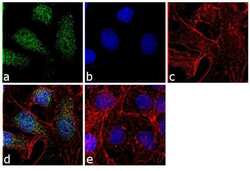
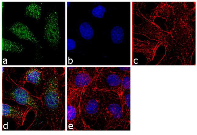
- Immunofluorescence analysis of Proteasome 20S Subunit alpha-5 was performed using 70% confluent log phase Hep G2 cells. The cells were fixed with 4% paraformaldehyde for 10 minutes, permeabilized with 0.1% Triton™ X-100 for 10 minutes, and blocked with 1% BSA for 1 hour at room temperature. The cells were labeled with PSMA5 Rabbit Polyclonal (Product # PA1-1962) at 2 µg/mL in 0.1% BSA and incubated for 3 hours at room temperature and then labeled with Goat anti-Rabbit IgG (H+L) Superclonal™ Secondary Antibody, Alexa Fluor® 488 conjugate (Product # A27034) at a dilution of 1:2000 for 45 minutes at room temperature (Panel a: green). Nuclei (Panel b: blue) were stained with SlowFade® Gold Antifade Mountant with DAPI (Product # S36938). F-actin (Panel c: red) was stained with Rhodamine Phalloidin (Product # R415, 1:300). Panel d represents the merged image showing cytoplasmic and nuclear localization. Panel e shows the no primary antibody control. The images were captured at 60X magnification.

- Submitted by
- Invitrogen Antibodies (provider)
- Main image

- Experimental details
- Immunofluorescence of HeLa cells using Product # PA1-1962.
- Submitted by
- Invitrogen Antibodies (provider)
- Main image
- Experimental details
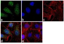
- Immunofluorescence analysis of Proteasome 20S Subunit alpha-5 was performed using 70% confluent log phase Hep G2 cells. The cells were fixed with 4% paraformaldehyde for 10 minutes, permeabilized with 0.1% Triton™ X-100 for 10 minutes, and blocked with 1% BSA for 1 hour at room temperature. The cells were labeled with PSMA5 Rabbit Polyclonal (Product # PA1-1962) at 2 µg/mL in 0.1% BSA and incubated for 3 hours at room temperature and then labeled with Goat anti-Rabbit IgG (Heavy Chain) Superclonal™ Secondary Antibody, Alexa Fluor® 488 conjugate (Product # A27034) at a dilution of 1:2000 for 45 minutes at room temperature (Panel a: green). Nuclei (Panel b: blue) were stained with SlowFade® Gold Antifade Mountant with DAPI (Product # S36938). F-actin (Panel c: red) was stained with Rhodamine Phalloidin (Product # R415, 1:300). Panel d represents the merged image showing cytoplasmic and nuclear localization. Panel e shows the no primary antibody control. The images were captured at 60X magnification.
Supportive validation
- Submitted by
- Invitrogen Antibodies (provider)
- Main image
- Experimental details
- NULL
- Submitted by
- Invitrogen Antibodies (provider)
- Main image
- Experimental details
- NULL

- Submitted by
- Invitrogen Antibodies (provider)
- Main image
- Experimental details
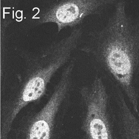
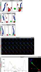
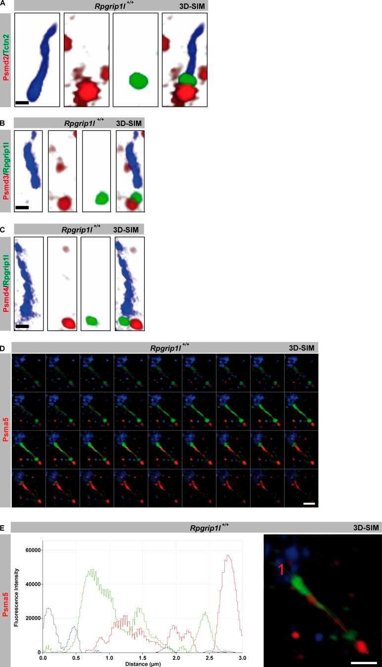
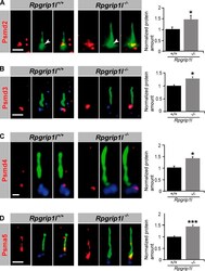
- Figure 6. Rpgrip1l deficiency results in an accumulation of proteasomal subunit components at the base of cilia. (A-D) Immunofluorescence on MEFs of E12.5 WT and Rpgrip1l -/- embryos (both genotypes: Psmd2, n = 7 embryos; Psmd3, n = 3 embryos; Psmd4, n = 3 embryos; Psma5, n = 5 embryos). At least 10 cilia per embryo were used for Psmd2, Psmd3, and Psmd4 quantification, respectively, and 20 cilia per embryo were used for Psma5 quantification. The ciliary axoneme is marked by acetylated alpha-tubulin (green). The BB is marked by Pcnt (green; white arrowheads; A) or gamma-tubulin (blue; B-D). Error bars show standard error of the mean. *, P < 0.05; ***, P < 0.001. Bars: (A, B, and D) 1 um; (C) 0.5 um.
