Explore
 Validate
 Learn
Learn Western blot
 Immunocytochemistry
Antibody data
- Antibody Data
- Antigen structure
- References [1]
- Comments [0]
- Validations
- Immunocytochemistry [1]
- Immunohistochemistry [1]
- Other assay [1]
Submit
Validation data
Reference
Comment
Report error
- Product number
- PA5-27940 - Provider product page

- Provider
- Invitrogen Antibodies
- Product name
- GNAI3 Polyclonal Antibody
- Antibody type
- Polyclonal
- Antigen
- Recombinant full-length protein
- Description
- Recommended positive controls: 293T, A431, H1299, HeLaS3, HepG2, Molt-4, Raji, mouse brain. Predicted reactivity: Mouse (97%), Rat (97%), Zebrafish (88%), Japanese Medaka (91%), Xenopus laevis (93%), Pig (99%), Chicken (97%), Rhesus Monkey (100%), Bovine (98%), Guinea pig (97%). Store product as a concentrated solution. Centrifuge briefly prior to opening the vial.
- Reactivity
- Human, Mouse
- Host
- Rabbit
- Isotype
- IgG
- Vial size
- 100 μL
- Concentration
- 0.94 mg/mL
- Storage
- Store at 4°C short term. For long term storage, store at -20°C, avoiding freeze/thaw cycles.
Submitted references Downregulation of GNAI3 Promotes the Pathogenesis of Methionine/Choline-Deficient Diet-Induced Nonalcoholic Fatty Liver Disease.
Zhu H, Ge K, Lu J, Jia C
Gut and liver 2020 Jul 15;14(4):492-499
Gut and liver 2020 Jul 15;14(4):492-499
No comments: Submit comment
Supportive validation
- Submitted by
- Invitrogen Antibodies (provider)
- Main image
- Experimental details
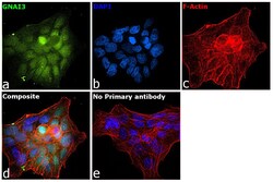
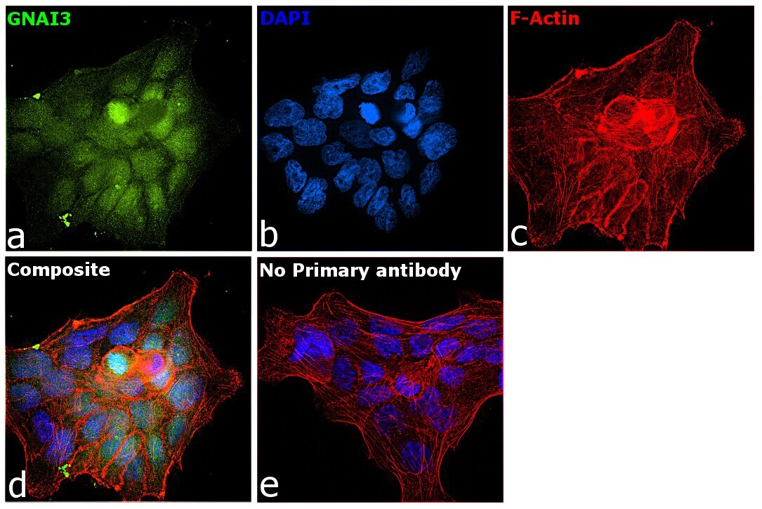
- Immunofluorescence analysis of GNAI3 was performed using 70% confluent log phase A-431 cells. The cells were fixed with 4% paraformaldehyde for 10 minutes, permeabilized with 0.1% Triton™ X-100 for 15 minutes, and blocked with 1% BSA for 1 hour at room temperature. The cells were labeled with GNAI3 Polyclonal Antibody (Product # PA5-27940) at 1:200 dilution in 0.1% BSA, incubated at 4 degree Celsius overnight and then labeled with Goat anti-Rabbit IgG (Heavy Chain), Superclonal™ Recombinant Secondary Antibody, Alexa Fluor 488 (Product # A27034) at a dilution of 1:2000 for 45 minutes at room temperature (Panel a: green). Nuclei (Panel b: blue) were stained with SlowFade® Gold Antifade Mountant with DAPI (Product # S36938). F-actin (Panel c: red) was stained with Rhodamine Phalloidin (Product # R415, 1:300). Panel d represents the merged image showing nuclear and cytoplasmic localization. Panel e represents control cells with no primary antibody to assess background. The images were captured at 60X magnification.
Supportive validation
- Submitted by
- Invitrogen Antibodies (provider)
- Main image
- Experimental details
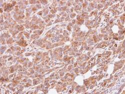
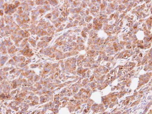
- Immunohistochemical analysis of paraffin-embedded SAS xenograft, using G protein alpha Inhibitor 3 (Product # PA5-27940) antibody at 1:500 dilution. Antigen Retrieval: Citrate buffer, pH 6.0, 15 min.
Supportive validation
- Submitted by
- Invitrogen Antibodies (provider)
- Main image

- Experimental details
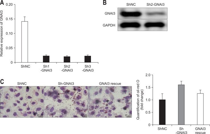
- Fig. 3 Construction of GNAI3 KD HepG2 cells and the effect of GNAI3 KD on cellular lipid accumulation. (A) Quantitative reverse transcription polymerase chain reaction analysis of GNAI3 mRNA expression in HepG2 cells transfected with lentiviral vectors carrying GNAI3 shRNA. (B) Western blotting analysis of GNAI3 protein expression levels in GNAI3 KD and control HepG2 cells. (C) Oil Red O staining of control, GNAI3 KD and rescued HepG2 cells after free fatty acid treatment. Positive staining for Oil Red O indicates intracellular lipid accumulation. Quantification of Oil Red O staining is shown in the right panel. The bar graphs show the mean+-standard error of the mean. p