Explore
 Validate
 Learn
Learn Western blot
 Blocking/Neutralizing
Antibody data
- Antibody Data
- Antigen structure
- References [6]
- Comments [0]
- Validations
- Blocking/Neutralizing [1]
Submit
Validation data
Reference
Comment
Report error
- Product number
- MAB4161 - Provider product page

- Provider
- Novus Biologicals
- Product name
- Rat Monoclonal M-CSF Antibody
- Antibody type
- Monoclonal
- Description
- Protein A or G purified from hybridoma culture supernatant. Detects mouse M-CSF in direct ELISAs and Western blots. In direct ELISAs and Western blots, no cross-reactivity with recombinant human M-CSF or recombinant mouse SCF is observed.
- Reactivity
- Mouse
- Host
- Rat
- Isotype
- IgG
- Vial size
- 500 ug
- Storage
- Use a manual defrost freezer and avoid repeated freeze-thaw cycles. 12 months from date of receipt, -20 to -70 degreesC as supplied. 1 month, 2 to 8 degreesC under sterile conditions after reconstitution. 6 months, -20 to -70 degreesC under sterile conditions after reconstitution.
Submitted references Bone-derived Nestin-positive mesenchymal stem cells improve cardiac function via recruiting cardiac endothelial cells after myocardial infarction.
Interleukin-18 Amplifies Macrophage Polarization and Morphological Alteration, Leading to Excessive Angiogenesis.
An inflammatory gene signature distinguishes neurofibroma Schwann cells and macrophages from cells in the normal peripheral nervous system.
CSF-1 receptor-mediated differentiation of a new type of monocytic cell with B cell-stimulating activity: its selective dependence on IL-34.
Colony-stimulating factor 1 receptor (CSF1R) signaling in injured neurons facilitates protection and survival.
Osteoblasts support B-lymphocyte commitment and differentiation from hematopoietic stem cells.
Lu D, Liao Y, Zhu SH, Chen QC, Xie DM, Liao JJ, Feng X, Jiang MH, He W
Stem cell research & therapy 2019 Apr 27;10(1):127
Stem cell research & therapy 2019 Apr 27;10(1):127
Interleukin-18 Amplifies Macrophage Polarization and Morphological Alteration, Leading to Excessive Angiogenesis.
Kobori T, Hamasaki S, Kitaura A, Yamazaki Y, Nishinaka T, Niwa A, Nakao S, Wake H, Mori S, Yoshino T, Nishibori M, Takahashi H
Frontiers in immunology 2018;9:334
Frontiers in immunology 2018;9:334
An inflammatory gene signature distinguishes neurofibroma Schwann cells and macrophages from cells in the normal peripheral nervous system.
Choi K, Komurov K, Fletcher JS, Jousma E, Cancelas JA, Wu J, Ratner N
Scientific reports 2017 Mar 3;7:43315
Scientific reports 2017 Mar 3;7:43315
CSF-1 receptor-mediated differentiation of a new type of monocytic cell with B cell-stimulating activity: its selective dependence on IL-34.
Yamane F, Nishikawa Y, Matsui K, Asakura M, Iwasaki E, Watanabe K, Tanimoto H, Sano H, Fujiwara Y, Stanley ER, Kanayama N, Mabbott NA, Magari M, Ohmori H
Journal of leukocyte biology 2014 Jan;95(1):19-31
Journal of leukocyte biology 2014 Jan;95(1):19-31
Colony-stimulating factor 1 receptor (CSF1R) signaling in injured neurons facilitates protection and survival.
Luo J, Elwood F, Britschgi M, Villeda S, Zhang H, Ding Z, Zhu L, Alabsi H, Getachew R, Narasimhan R, Wabl R, Fainberg N, James ML, Wong G, Relton J, Gambhir SS, Pollard JW, Wyss-Coray T
The Journal of experimental medicine 2013 Jan 14;210(1):157-72
The Journal of experimental medicine 2013 Jan 14;210(1):157-72
Osteoblasts support B-lymphocyte commitment and differentiation from hematopoietic stem cells.
Zhu J, Garrett R, Jung Y, Zhang Y, Kim N, Wang J, Joe GJ, Hexner E, Choi Y, Taichman RS, Emerson SG
Blood 2007 May 1;109(9):3706-12
Blood 2007 May 1;109(9):3706-12
No comments: Submit comment
Supportive validation
- Submitted by
- Novus Biologicals (provider)
- Main image
- Experimental details
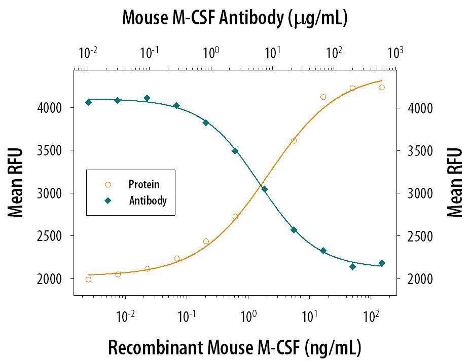
- Cell Proliferation Induced by M-CSF and Neutralization by Mouse M-CSF Antibody. Recombinant Mouse M-CSF (Catalog # 416-ML) stimulates proliferation in the M-NFS-60 mouse myelogenous luekemia lymphoblast cell line in a dose-dependent manner (orange line). Proliferation elicited by Recombinant Mouse M-CSF (10 ng/mL) is neutralized (green line) by increasing concentrations of Mouse M-CSF Monoclonal Antibody (Catalog # MAB4161). The ND50 is typically 3-12 µg/mL.