Explore
 Validate
 Learn
Learn Western blot
Antibody data
- Antibody Data
- Antigen structure
- References [0]
- Comments [0]
- Validations
- Western blot [1]
- Immunocytochemistry [7]
- Immunoprecipitation [1]
- Immunohistochemistry [5]
- Other assay [1]
Submit
Validation data
Reference
Comment
Report error
- Product number
- MA5-32139 - Provider product page

- Provider
- Invitrogen Antibodies
- Product name
- M-CSF Recombinant Rabbit Monoclonal Antibody (SU0413)
- Antibody type
- Monoclonal
- Antigen
- Synthetic peptide
- Description
- Recombinant rabbit monoclonal antibodies are produced using in vitro expression systems. The expression systems are developed by cloning in the specific antibody DNA sequences from immunoreactive rabbits. Then, individual clones are screened to select the best candidates for production. The advantages of using recombinant rabbit monoclonal antibodies include: better specificity and sensitivity, lot-to-lot consistency, animal origin-free formulations, and broader immunoreactivity to diverse targets due to larger rabbit immune repertoire.
- Reactivity
- Human
- Host
- Rabbit
- Isotype
- IgG
- Antibody clone number
- SU0413
- Vial size
- 100 μL
- Concentration
- 1 mg/mL
- Storage
- Store at 4°C short term. For long term storage, store at -20°C, avoiding freeze/thaw cycles.
No comments: Submit comment
Supportive validation
- Submitted by
- Invitrogen Antibodies (provider)
- Main image
- Experimental details
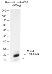
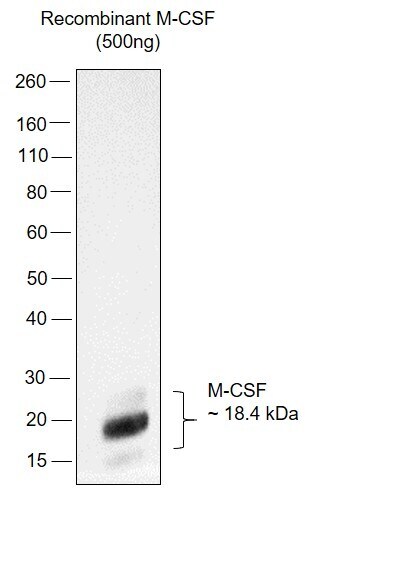
- Western Blot was performed using Anti-M-CSF Recombinant Rabbit Monoclonal Antibody (SU0413) (Product # MA5-32139) and an 18.5 kDa band corresponding to Macrophage colony-stimulating factor 1 was observed in recombinant M-CSF protein. Recombinant M-CSF (500 ng µg lysate) was electrophoresed using NuPAGE™ 4-12% Bis-Tris Protein Gel (Product # NP0322BOX). Resolved proteins were then transferred onto a Nitrocellulose membrane (Product # IB23002) by iBlot® 2 Dry Blotting System (Product # IB21001). The Blot was probed with the primary antibody (1:1000 dilution) and detected by chemiluminescence with Goat anti-Rabbit IgG (Heavy Chain) Superclonal™ Recombinant Secondary Antibody, HRP (Product # A27036, 1:4000 dilution) using the iBright FL 1000 (Product # A32752). Chemiluminescent detection was performed using Novex® ECL Chemiluminescent Substrate Reagent Kit (Product # WP20005).
Supportive validation
- Submitted by
- Invitrogen Antibodies (provider)
- Main image
- Experimental details
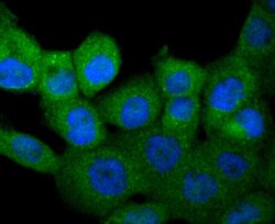
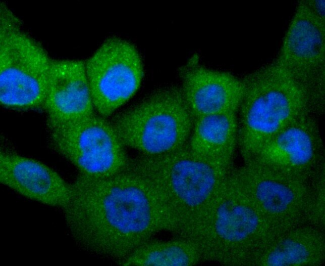
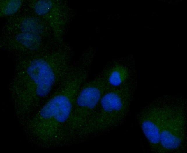
- Immunocytochemical analysis of M-CSF in A431 cells using a M-CSF Monoclonal antibody (Product # MA5-32139) as seen in green. The nuclear counter stain is DAPI (blue). Cells were fixed in paraformaldehyde, permeabilised with 0.25% Triton X100/PBS.
- Submitted by
- Invitrogen Antibodies (provider)
- Main image

- Experimental details
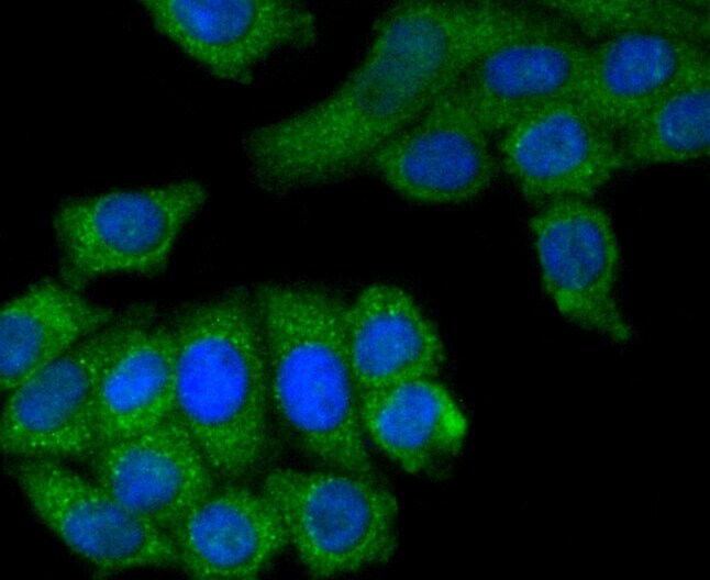
- Immunocytochemical analysis of M-CSF in HepG2 cells using a M-CSF Monoclonal antibody (Product # MA5-32139) as seen in green. The nuclear counter stain is DAPI (blue). Cells were fixed in paraformaldehyde, permeabilised with 0.25% Triton X100/PBS.
- Submitted by
- Invitrogen Antibodies (provider)
- Main image

- Experimental details
- Immunocytochemical analysis of M-CSF in Hela cells using a M-CSF Monoclonal antibody (Product # MA5-32139) as seen in green. The nuclear counter stain is DAPI (blue). Cells were fixed in paraformaldehyde, permeabilised with 0.25% Triton X100/PBS.

- Submitted by
- Invitrogen Antibodies (provider)
- Main image
- Experimental details
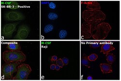
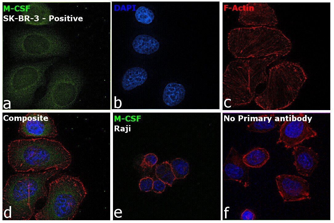
- Immunofluorescence analysis of Macrophage colony-stimulating factor 1 was performed using 70% confluent log phase SK-BR-3 and Raji cells. The cells were fixed with 4% paraformaldehyde for 10 minutes, permeabilized with 0.1% Triton™ X-100 for 15 minutes, and blocked with 2% BSA for 45 minutes at room temperature. The cells were labeled with M-CSF Recombinant Rabbit Monoclonal Antibody (SU0413) (Product # MA5-32139) at 1:100 in 0.1% BSA, incubated at 4 degree celsius overnight, and then labeled with Donkey anti-Rabbit IgG (H+L) Highly Cross-Adsorbed Secondary Antibody, Alexa Fluor Plus 488 (Product # A32790), (1:2000 dilution), for 45 minutes at room temperature (Panel a: Green). Nuclei (Panel b:Blue) were stained with ProLong™ Diamond Antifade Mountant with DAPI (Product # P36962). F-actin (Panel c: Red) was stained with Rhodamine Phalloidin (Product # R415, 1:300). Panel d represents the merged image showing Cytoplasmic localization in SK-BR-3 but not in Raji cells which are reported to be low or negative for M-CSF expression. Panel e represents control cells with no primary antibody to assess background. The images were captured at 60X magnification.

- Submitted by
- Invitrogen Antibodies (provider)
- Main image
- Experimental details
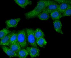
- Immunocytochemical analysis of M-CSF in HepG2 cells using a M-CSF Monoclonal antibody (Product # MA5-32139) as seen in green. The nuclear counter stain is DAPI (blue). Cells were fixed in paraformaldehyde, permeabilised with 0.25% Triton X100/PBS.

- Submitted by
- Invitrogen Antibodies (provider)
- Main image

- Experimental details
- Immunocytochemical analysis of M-CSF in Hela cells using a M-CSF Monoclonal antibody (Product # MA5-32139) as seen in green. The nuclear counter stain is DAPI (blue). Cells were fixed in paraformaldehyde, permeabilised with 0.25% Triton X100/PBS.
- Submitted by
- Invitrogen Antibodies (provider)
- Main image

- Experimental details
- Immunofluorescence analysis of Macrophage colony-stimulating factor 1 was performed using 70% confluent log phase SK-BR-3 and Raji cells. The cells were fixed with 4% paraformaldehyde for 10 minutes, permeabilized with 0.1% Triton™ X-100 for 15 minutes, and blocked with 2% BSA for 45 minutes at room temperature. The cells were labeled with M-CSF Recombinant Rabbit Monoclonal Antibody (SU0413) (Product # MA5-32139) at 1:100 in 0.1% BSA, incubated at 4 degree celsius overnight, and then labeled with Donkey anti-Rabbit IgG (H+L) Highly Cross-Adsorbed Secondary Antibody, Alexa Fluor Plus 488 (Product # A32790), (1:2000 dilution), for 45 minutes at room temperature (Panel a: Green). Nuclei (Panel b:Blue) were stained with ProLong™ Diamond Antifade Mountant with DAPI (Product # P36962). F-actin (Panel c: Red) was stained with Rhodamine Phalloidin (Product # R415, 1:300). Panel d represents the merged image showing Cytoplasmic localization in SK-BR-3 but not in Raji cells which are reported to be low or negative for M-CSF expression. Panel e represents control cells with no primary antibody to assess background. The images were captured at 60X magnification.
Supportive validation
- Submitted by
- Invitrogen Antibodies (provider)
- Main image

- Experimental details
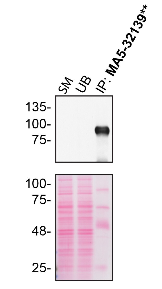
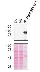
- U87-MG cells were washed 3x with PBS and starved for ~18 hrs. Immunoprecipitation of CSF1 was performed on U87-MG concentrated cell culture media, concentrated by centrifuging for 10 min at 500 x g to eliminate cells and larger contaminants, then for 10 min at 4500 x g to eliminate smaller contaminants. Culture media were concentrated using a centrifugal filter with a membrane NMWL of 10 kDa at 4000 x g for 10 min. Antibody-bead conjugates were prepared by adding 2 µg of CSF1 recombinant monoclonal antibody (Product # MA5-32139) with 30 µL of protein A-Sepharose beads and rocked overnight at 4 deg. 400 µg of lysate was incubated with an antibody-bead conjugate for 2 hours at 4 deg. Following centrifugation and multiple washes, 10% starting material (SM), 10% unbound fraction (UB) and immunoprecipitated fraction (IP) were processed for immunoblot using another CSF1 antibody. Ponceau stained transfer of blot is shown. Data courtesy of YCharOS Inc., an open science company with the mission of characterizing commercially available antibodies using knockout validation.

Supportive validation
- Submitted by
- Invitrogen Antibodies (provider)
- Main image
- Experimental details
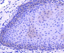
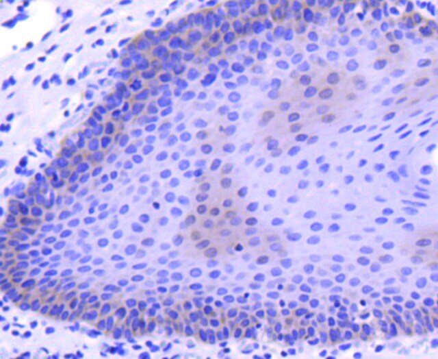
- Immunohistochemical analysis of M-CSF of paraffin-embedded Human tonsil tissue using a M-CSF Monoclonal antibody (Product #MA5-32139). Counter stained with hematoxylin.
- Submitted by
- Invitrogen Antibodies (provider)
- Main image
- Experimental details
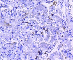
- Immunohistochemical analysis of M-CSF of paraffin-embedded Human lung tissue using a M-CSF Monoclonal antibody (Product #MA5-32139). Counter stained with hematoxylin.

- Submitted by
- Invitrogen Antibodies (provider)
- Main image
- Experimental details
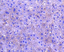
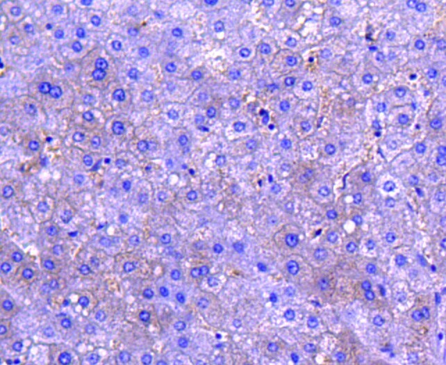
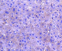
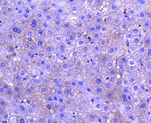
- Immunohistochemical analysis of M-CSF of paraffin-embedded Human liver tissue using a M-CSF Monoclonal antibody (Product #MA5-32139). Counter stained with hematoxylin.
- Submitted by
- Invitrogen Antibodies (provider)
- Main image

- Experimental details
- Immunohistochemical analysis of M-CSF of paraffin-embedded Human lung tissue using a M-CSF Monoclonal antibody (Product #MA5-32139). Counter stained with hematoxylin.

- Submitted by
- Invitrogen Antibodies (provider)
- Main image
- Experimental details
- Immunohistochemical analysis of M-CSF of paraffin-embedded Human liver tissue using a M-CSF Monoclonal antibody (Product #MA5-32139). Counter stained with hematoxylin.
Supportive validation
- Submitted by
- Invitrogen Antibodies (provider)
- Main image
- Experimental details
- U87-MG cells were washed 3x with PBS and starved for ~18 hrs. Immunoprecipitation of CSF1 was performed on U87-MG concentrated cell culture media, concentrated by centrifuging for 10 min at 500 x g to eliminate cells and larger contaminants, then for 10 min at 4500 x g to eliminate smaller contaminants. Culture media were concentrated using a centrifugal filter with a membrane NMWL of 10 kDa at 4000 x g for 10 min. Antibody-bead conjugates were prepared by adding 2 µg of CSF1 recombinant monoclonal antibody (Product # MA5-32139) with 30 µL of protein A-Sepharose beads and rocked overnight at 4 deg. 400 µg of lysate was incubated with an antibody-bead conjugate for 2 hours at 4 deg. Following centrifugation and multiple washes, 10% starting material (SM), 10% unbound fraction (UB) and immunoprecipitated fraction (IP) were processed for immunoblot using another CSF1 antibody. Ponceau stained transfer of blot is shown. Data courtesy of YCharOS Inc., an open science company with the mission of characterizing commercially available antibodies using knockout validation.