Explore
 Validate
 Learn
Learn Western blot
 Immunohistochemistry
Antibody data
- Antibody Data
- Antigen structure
- References [3]
- Comments [0]
- Validations
- Immunohistochemistry [1]
- Flow cytometry [1]
Submit
Validation data
Reference
Comment
Report error
- Product number
- AF3970 - Provider product page

- Provider
- R&D Systems
- Product name
- Mouse/Rat DLL1 Antibody
- Antibody type
- Polyclonal
- Description
- Antigen Affinity-purified. Detects rat and mouse DLL1 in direct ELISAs and Western blots. In direct ELISAs, approximately 50% cross-reactivity with recombinant human DLL-1 is observed.
- Reactivity
- Mouse, Rat
- Host
- Sheep
- Conjugate
- Unconjugated
- Antigen sequence
P97677- Isotype
- IgG
- Vial size
- 100 ug
- Concentration
- LYOPH
- Storage
- Use a manual defrost freezer and avoid repeated freeze-thaw cycles. 12 months from date of receipt, -20 to -70 °C as supplied. 1 month, 2 to 8 °C under sterile conditions after reconstitution. 6 months, -20 to -70 °C under sterile conditions after reconstitution.
Submitted references MPDZ promotes DLL4-induced Notch signaling during angiogenesis.
Cadherin-based adhesions in the apical endfoot are required for active Notch signaling to control neurogenesis in vertebrates.
Single-cell gene profiling defines differential progenitor subclasses in mammalian neurogenesis.
Tetzlaff F, Adam MG, Feldner A, Moll I, Menuchin A, Rodriguez-Vita J, Sprinzak D, Fischer A
eLife 2018 Apr 5;7
eLife 2018 Apr 5;7
Cadherin-based adhesions in the apical endfoot are required for active Notch signaling to control neurogenesis in vertebrates.
Hatakeyama J, Wakamatsu Y, Nagafuchi A, Kageyama R, Shigemoto R, Shimamura K
Development (Cambridge, England) 2014 Apr;141(8):1671-82
Development (Cambridge, England) 2014 Apr;141(8):1671-82
Single-cell gene profiling defines differential progenitor subclasses in mammalian neurogenesis.
Kawaguchi A, Ikawa T, Kasukawa T, Ueda HR, Kurimoto K, Saitou M, Matsuzaki F
Development (Cambridge, England) 2008 Sep;135(18):3113-24
Development (Cambridge, England) 2008 Sep;135(18):3113-24
No comments: Submit comment
Supportive validation
- Submitted by
- R&D Systems (provider)
- Main image

- Experimental details
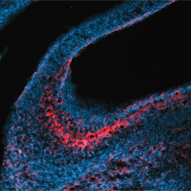
- DLL1 in Embryonic Mouse Stomach. DLL1 was detected in immersion fixed frozen sections of embryonic mouse stomach (E13.5) using 10 µg/mL Sheep Anti-Mouse/Rat DLL1 Antigen Affinity-purified Polyclonal Antibody (Catalog # AF3970) overnight at 4 °C. Tissue was stained with the NorthernLights™ 557-conjugated Anti-Sheep IgG Secondary Antibody (red; Catalog # NL010) and counterstained with DAPI (blue). View our protocol for Fluorescent IHC Staining of Frozen Tissue Sections.
Supportive validation
- Submitted by
- R&D Systems (provider)
- Main image

- Experimental details
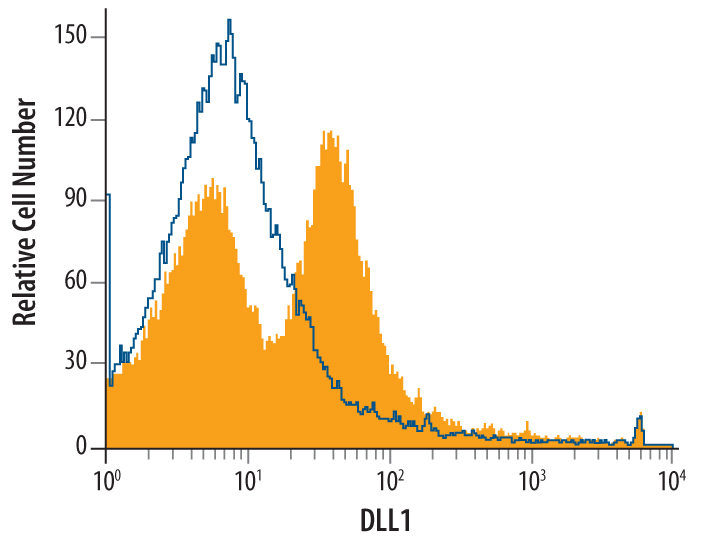
- Detection of DLL1 in Mouse Splenocytes by Flow Cytometry. Mouse splenocytes were stained with Sheep Anti-Mouse/Rat DLL1 Antigen Affinity-purified Polyclonal Antibody (Catalog # AF3970, filled histogram) or control antibody (Catalog # 5-001-A, open histogram), followed by NorthernLights™ 637-conjugated Anti-Sheep IgG Secondary Antibody (Catalog # NL011).