Explore
 Validate
 Learn
Learn Western blot
 Immunocytochemistry
Antibody data
- Antibody Data
- Antigen structure
- References [4]
- Comments [0]
- Validations
- Western blot [2]
- Immunohistochemistry [4]
Submit
Validation data
Reference
Comment
Report error
- Product number
- NBP1-88921 - Provider product page

- Provider
- Novus Biologicals
- Proper citation
- Novus Cat#NBP1-88921, RRID:AB_11005096
- Product name
- Rabbit Polyclonal LPCAT2 Antibody
- Antibody type
- Polyclonal
- Description
- Immunogen affinity purified. Specificity of human, rat LPCAT2 antibody verified on a Protein Array containing target protein plus 383 other non-specific proteins.
- Reactivity
- Human, Mouse, Rat
- Host
- Rabbit
- Isotype
- IgG
- Vial size
- 0.1 ml
- Storage
- Store at 4C short term. Aliquot and store at -20C long term. Avoid freeze-thaw cycles.
Submitted references ROBO4 deletion ameliorates PAF-mediated skin inflammation via regulating the mRNA translation efficiency of LPCAT1/LPCAT2 and the expression of PAF receptor.
Lipidome-based rapid diagnosis with machine learning for detection of TGF-β signalling activated area in head and neck cancer.
The expression of Toll-like receptor 4, 7 and co-receptors in neurochemical sub-populations of rat trigeminal ganglion sensory neurons.
Immunofluorescence and fluorescent-protein tagging show high correlation for protein localization in mammalian cells.
Xiao X, Zhuang X, Xu C, Chen H, Zhu W, Pang C, Zhang M
International journal of biological sciences 2020;16(6):1086-1095
International journal of biological sciences 2020;16(6):1086-1095
Lipidome-based rapid diagnosis with machine learning for detection of TGF-β signalling activated area in head and neck cancer.
Ishii H, Saitoh M, Sakamoto K, Sakamoto K, Saigusa D, Kasai H, Ashizawa K, Miyazawa K, Takeda S, Masuyama K, Yoshimura K
British journal of cancer 2020 Mar;122(7):995-1004
British journal of cancer 2020 Mar;122(7):995-1004
The expression of Toll-like receptor 4, 7 and co-receptors in neurochemical sub-populations of rat trigeminal ganglion sensory neurons.
Helley MP, Abate W, Jackson SK, Bennett JH, Thompson SW
Neuroscience 2015 Dec 3;310:686-98
Neuroscience 2015 Dec 3;310:686-98
Immunofluorescence and fluorescent-protein tagging show high correlation for protein localization in mammalian cells.
Stadler C, Rexhepaj E, Singan VR, Murphy RF, Pepperkok R, Uhlén M, Simpson JC, Lundberg E
Nature methods 2013 Apr;10(4):315-23
Nature methods 2013 Apr;10(4):315-23
No comments: Submit comment
Supportive validation
- Submitted by
- Novus Biologicals (provider)
- Main image
- Experimental details
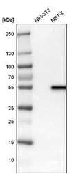
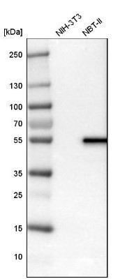
- Western Blot: LPCAT2 Antibody [NBP1-88921] - Analysis in mouse cell line NIH-3T3 and rat cell line NBT-II.
- Submitted by
- Novus Biologicals (provider)
- Main image
- Experimental details
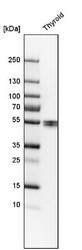
- Western Blot: LPCAT2 Antibody [NBP1-88921] - Analysis in human thyroid gland tissue.

Supportive validation
- Submitted by
- Novus Biologicals (provider)
- Main image
- Experimental details
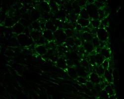
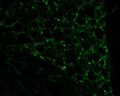
- Immunohistochemistry-Frozen: LPCAT2 Antibody [NBP1-88921] - Staining of LPCAT2 in rat neuronal tissue using anti-LPCAT2 antibody. Image from verified customer review.
- Submitted by
- Novus Biologicals (provider)
- Main image
- Experimental details
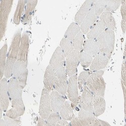
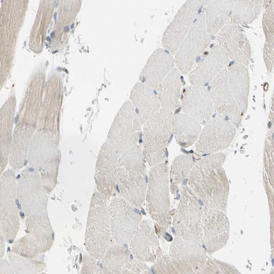
- Immunohistochemistry-Paraffin: LPCAT2 Antibody [NBP1-88921] - Staining of human skeletal muscle shows low expression as expected.
- Submitted by
- Novus Biologicals (provider)
- Main image

- Experimental details
- Immunohistochemistry-Paraffin: LPCAT2 Antibody [NBP1-88921] - Staining of human thyroid gland shows high expression.
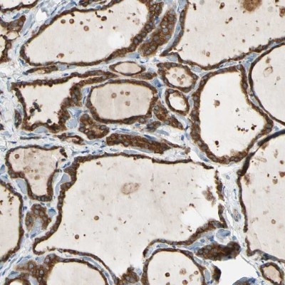
- Submitted by
- Novus Biologicals (provider)
- Main image
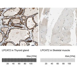
- Experimental details
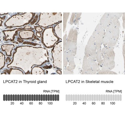
- Immunohistochemistry-Paraffin: LPCAT2 Antibody [NBP1-88921] - Staining in human thyroid gland and skeletal muscle tissues using anti-LPCAT2 antibody. Corresponding LPCAT2 RNA-seq data are presented for the same tissues.