 Explore
 Validate
 Learn
Learn Western blot
 Immunocytochemistry
Antibody data
- Antibody Data
- Antigen structure
- References [1]
- Comments [0]
- Validations
- Immunocytochemistry [2]
- Immunohistochemistry [3]
Submit
Validation data
Reference
Comment
Report error
- Product number
- 710888 - Provider product page

- Provider
- Invitrogen Antibodies
- Product name
- WNT2B Recombinant Superclonal™ Antibody (17HCLC)
- Antibody type
- Other
- Antigen
- Synthetic peptide
- Description
- This antibody is predicted to react with Monkey, Pig and Rabbit. Recombinant rabbit Superclonal™ antibodies are unique offerings from Thermo Fisher Scientific. They are comprised of a selection of multiple different recombinant monoclonal antibodies, providing the best of both worlds - the sensitivity of polyclonal antibodies with the specificity of monoclonal antibodies - all delivered with the consistency only found in a recombinant antibody. While functionally the same as a polyclonal antibody - recognizing multiple epitope sites on the target and producing higher detection sensitivity for low abundance targets - a recombinant rabbit Superclonal™ antibody has a known mixture of light and heavy chains. The exact population can be produced in every lot, circumventing the biological variability typically associated with polyclonal antibody production. Note: Formerly called “Recombinant polyclonal antibody”, this product is now rebranded as “Recombinant Superclonal™ antibody”. The physical product and the performance remain unchanged.
- Reactivity
- Human, Mouse
- Host
- Rabbit
- Isotype
- IgG
- Antibody clone number
- 17HCLC
- Vial size
- 100 μg
- Concentration
- 0.5 mg/mL
- Storage
- Store at 4°C short term. For long term storage, store at -20°C, avoiding freeze/thaw cycles.
Submitted references Noggin regulates foregut progenitor cell programming, and misexpression leads to esophageal atresia.
Pinzon-Guzman C, Sangadala S, Riera KM, Popova EY, Manning E, Huh WJ, Alexander MS, Shelton JS, Boden SD, Goldenring JR
The Journal of clinical investigation 2020 Aug 3;130(8):4396-4410
The Journal of clinical investigation 2020 Aug 3;130(8):4396-4410
No comments: Submit comment
Supportive validation
- Submitted by
- Invitrogen Antibodies (provider)
- Main image

- Experimental details
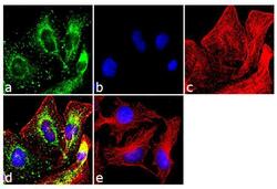
- Immunofluorescence was performed on methanol fixed MDA-MB-231 cells for detection of WNT2B/13 using Anti-WNT2B/13 Recombinant Rabbit Monoclonal Antibody (Product # 701856, 2 µg/mL), alpha-Tubulin Monoclonal Antibody (Product # 32-2500, 1 µg/mL) and labeled with Goat anti-Rabbit IgG (H+L) Superclonal™ Secondary Antibody, Alexa Fluor® 488 conjugate (Product # A27034, 1:2000), Goat anti-Mouse IgG Secondary Antibody, Alexa Fluor® 594conjugate (Product # A-11032, 1:400) respectively. Panel a) shows representative cells that were stained for detection and localization of WNT2B/13 protein (green), Panel b) is stained for nuclei (blue) using SlowFade® Gold Antifade Mountant with DAPI (Product # S36938,). Panel c) represents cytoskeletal alpha-tubulin staining (red). Panel d) is a composite image of Panels a, b and c clearly demonstrating cytoplasmic localization of WNT2B/13. Panel e) represents control cells with no primary antibody to assess background.

- Submitted by
- Invitrogen Antibodies (provider)
- Main image
- Experimental details
- Immunofluorescence was performed on methanol fixed MDA-MB-231 cells for detection of WNT2B/13 using Anti-WNT2B/13 Recombinant Rabbit Monoclonal Antibody (Product # 701856, 2 µg/mL), alpha-Tubulin Monoclonal Antibody (Product # 32-2500, 1 µg/mL) and labeled with Goat anti-Rabbit IgG (Heavy Chain) Superclonal™ Secondary Antibody, Alexa Fluor® 488 conjugate (Product # A27034, 1:2000), Goat anti-Mouse IgG Secondary Antibody, Alexa Fluor® 594conjugate (Product # A-11032, 1:400) respectively. Panel a) shows representative cells that were stained for detection and localization of WNT2B/13 protein (green), Panel b) is stained for nuclei (blue) using SlowFade® Gold Antifade Mountant with DAPI (Product # S36938,). Panel c) represents cytoskeletal alpha-tubulin staining (red). Panel d) is a composite image of Panels a, b and c clearly demonstrating cytoplasmic localization of WNT2B/13. Panel e) represents control cells with no primary antibody to assess background.
Supportive validation
- Submitted by
- Invitrogen Antibodies (provider)
- Main image
- Experimental details
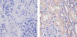
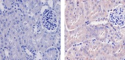
- Immunohistochemistry analysis of Wnt 2B/13 showing staining in the cytoplasm and nucleus of paraffin-embedded human kidney tissue (right) compared to a negative control without primary antibody (left). To expose target proteins, antigen retrieval was performed using 10mM sodium citrate (pH 6.0), microwaved for 8-15 min. Following antigen retrieval, tissues were blocked in 3% H2O2-methanol for 15 min at room temperature, washed with ddH2O and PBS, and then probed with a Wnt 2B/13 Recombinant Rabbit Superclonal™ Antibody (Product # 710888) diluted in 3% BSA-PBS at a dilution of 1:20 for 1 hour at 37ºC in a humidified chamber. Tissues were washed extensively in PBST and detection was performed using an HRP-conjugated secondary antibody followed by colorimetric detection using a DAB kit. Tissues were counterstained with hematoxylin and dehydrated with ethanol and xylene to prep for mounting.
- Submitted by
- Invitrogen Antibodies (provider)
- Main image
- Experimental details
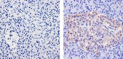
- Immunohistochemistry analysis of Wnt 2B/13 showing staining in the cytoplasm and nucleus of paraffin-embedded human pancreas tissue (right) compared to a negative control without primary antibody (left). To expose target proteins, antigen retrieval was performed using 10mM sodium citrate (pH 6.0), microwaved for 8-15 min. Following antigen retrieval, tissues were blocked in 3% H2O2-methanol for 15 min at room temperature, washed with ddH2O and PBS, and then probed with a Wnt 2B/13 Recombinant Rabbit Superclonal™ Antibody (Product # 710888) diluted in 3% BSA-PBS at a dilution of 1:20 for 1 hour at 37ºC in a humidified chamber. Tissues were washed extensively in PBST and detection was performed using an HRP-conjugated secondary antibody followed by colorimetric detection using a DAB kit. Tissues were counterstained with hematoxylin and dehydrated with ethanol and xylene to prep for mounting.

- Submitted by
- Invitrogen Antibodies (provider)
- Main image
- Experimental details
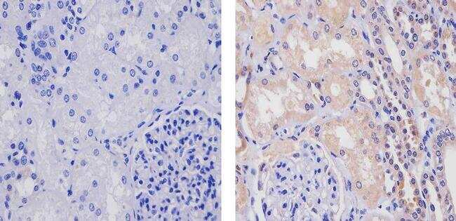
- Immunohistochemistry analysis of Wnt 2B/13 showing staining in the cytoplasm and nucleus of paraffin-embedded mouse kidney tissue (right) compared to a negative control without primary antibody (left). To expose target proteins, antigen retrieval was performed using 10mM sodium citrate (pH 6.0), microwaved for 8-15 min. Following antigen retrieval, tissues were blocked in 3% H2O2-methanol for 15 min at room temperature, washed with ddH2O and PBS, and then probed with a Wnt 2B/13 Recombinant Rabbit Superclonal™ Antibody (Product # 710888) diluted in 3% BSA-PBS at a dilution of 1:20 for 1 hour at 37ºC in a humidified chamber. Tissues were washed extensively in PBST and detection was performed using an HRP-conjugated secondary antibody followed by colorimetric detection using a DAB kit. Tissues were counterstained with hematoxylin and dehydrated with ethanol and xylene to prep for mounting.
