Explore
 Validate
 Learn
Learn Western blot
 Immunocytochemistry
Antibody data
- Antibody Data
- Antigen structure
- References [16]
- Comments [0]
- Validations
- Western blot [2]
- Immunocytochemistry [1]
Submit
Validation data
Reference
Comment
Report error
- Product number
- HPA025235 - Provider product page

- Provider
- Atlas Antibodies
- Proper citation
- Atlas Antibodies Cat#HPA025235, RRID:AB_1858718
- Product name
- Anti-VANGL1
- Antibody type
- Polyclonal
- Description
- Polyclonal Antibody against Human VANGL1, Gene description: VANGL planar cell polarity protein 1, Alternative Gene Names: STB2, Validated applications: ICC, WB, Uniprot ID: Q8TAA9, Storage: Store at +4°C for short term storage. Long time storage is recommended at -20°C.
- Reactivity
- Human
- Host
- Rabbit
- Conjugate
- Unconjugated
- Isotype
- IgG
- Vial size
- 100 µl
- Concentration
- 0.2 mg/ml
- Storage
- Store at +4°C for short term storage. Long time storage is recommended at -20°C.
- Handling
- The antibody solution should be gently mixed before use.
Submitted references Junctional Adhesion Molecule 3 Expression in the Mouse Airway Epithelium Is Linked to Multiciliated Cells
nNOS regulates ciliated cell polarity, ciliary beat frequency, and directional flow in mouse trachea
Planar cell polarity induces local microtubule bundling for coordinated ciliary beating
New mouse models for high resolution and live imaging of planar cell polarity proteins in vivo
The molecular dynamics of subdistal appendages in multi-ciliated cells.
A human ciliopathy reveals essential functions for NEK10 in airway mucociliary clearance
Multi-scale spatial heterogeneity enhances particle clearance in airway ciliary arrays
Non-canonical Wnt signalling regulates scarring in biliary disease via the planar cell polarity receptors
Polarized cellular mechanoresponse system for maintaining radial size in developing epithelial tubes
Domineering non-autonomy in Vangl1;Vangl2 double mutants demonstrates intercellular PCP signaling in the vertebrate inner ear
Long-term preservation of planar cell polarity in reversed tracheal epithelium.
Disruption of Core Planar Cell Polarity Signaling Regulates Renal Tubule Morphogenesis but Is Not Cystogenic
Observing planar cell polarity in multiciliated mouse airway epithelial cells
Flattop regulates basal body docking and positioning in mono- and multiciliated cells
Postnatal Refinement of Auditory Hair Cell Planar Polarity Deficits Occurs in the Absence of Vangl2
Planar cell polarity effector gene Intu regulates cell fate-specific differentiation of keratinocytes through the primary cilia
Mateos-Quiros C, Garrido-Jimenez S, Álvarez-Hernán G, Diaz-Chamorro S, Barrera-Lopez J, Francisco-Morcillo J, Roman A, Centeno F, Carvajal-Gonzalez J
Frontiers in Cell and Developmental Biology 2021;9
Frontiers in Cell and Developmental Biology 2021;9
nNOS regulates ciliated cell polarity, ciliary beat frequency, and directional flow in mouse trachea
Mikhailik A, Michurina T, Dikranian K, Hearn S, Maxakov V, Siller S, Takemaru K, Enikolopov G, Peunova N
Life Science Alliance 2021;4(5):e202000981
Life Science Alliance 2021;4(5):e202000981
Planar cell polarity induces local microtubule bundling for coordinated ciliary beating
Nakayama S, Yano T, Namba T, Konishi S, Takagishi M, Herawati E, Nishida T, Imoto Y, Ishihara S, Takahashi M, Furuta K, Oiwa K, Tamura A, Tsukita S
Journal of Cell Biology 2021;220(7)
Journal of Cell Biology 2021;220(7)
New mouse models for high resolution and live imaging of planar cell polarity proteins in vivo
Basta L, Hill-Oliva M, Paramore S, Sharan R, Goh A, Biswas A, Cortez M, Little K, Posfai E, Devenport D
Development 2021;148(18)
Development 2021;148(18)
The molecular dynamics of subdistal appendages in multi-ciliated cells.
Ryu H, Lee H, Lee J, Noh H, Shin M, Kumar V, Hong S, Kim J, Park S
Nature communications 2021 Jan 27;12(1):612
Nature communications 2021 Jan 27;12(1):612
A human ciliopathy reveals essential functions for NEK10 in airway mucociliary clearance
Chivukula R, Montoro D, Leung H, Yang J, Shamseldin H, Taylor M, Dougherty G, Zariwala M, Carson J, Daniels M, Sears P, Black K, Hariri L, Almogarri I, Frenkel E, Vinarsky V, Omran H, Knowles M, Tearney G, Alkuraya F, Sabatini D
Nature Medicine 2020;26(2):244-251
Nature Medicine 2020;26(2):244-251
Multi-scale spatial heterogeneity enhances particle clearance in airway ciliary arrays
Ramirez-San Juan G, Mathijssen A, He M, Jan L, Marshall W, Prakash M
Nature Physics 2020;16(9):958-964
Nature Physics 2020;16(9):958-964
Non-canonical Wnt signalling regulates scarring in biliary disease via the planar cell polarity receptors
Wilson D, Jarman E, Mellin R, Wilson M, Waddell S, Tsokkou P, Younger N, Raven A, Bhalla S, Noll A, Olde Damink S, Schaap F, Chen P, Bates D, Banales J, Dean C, Henderson D, Sansom O, Kendall T, Boulter L
Nature Communications 2020;11(1)
Nature Communications 2020;11(1)
Polarized cellular mechanoresponse system for maintaining radial size in developing epithelial tubes
Hirashima T, Adachi T
Development 2019
Development 2019
Domineering non-autonomy in Vangl1;Vangl2 double mutants demonstrates intercellular PCP signaling in the vertebrate inner ear
Stoller M, Roman O, Deans M
Developmental Biology 2018;437(1):17-26
Developmental Biology 2018;437(1):17-26
Long-term preservation of planar cell polarity in reversed tracheal epithelium.
Tsuji T, Nakamura R, Katsuno T, Kishimoto Y, Suehiro A, Yamashita M, Uozumi R, Nakamura T, Tateya I, Omori K
Respiratory research 2018 Feb 2;19(1):22
Respiratory research 2018 Feb 2;19(1):22
Disruption of Core Planar Cell Polarity Signaling Regulates Renal Tubule Morphogenesis but Is Not Cystogenic
Kunimoto K, Bayly R, Vladar E, Vonderfecht T, Gallagher A, Axelrod J
Current Biology 2017;27(20):3120-3131.e4
Current Biology 2017;27(20):3120-3131.e4
Observing planar cell polarity in multiciliated mouse airway epithelial cells
Vladar E, Lee Y, Stearns T, Axelrod J
2015;127
2015;127
Flattop regulates basal body docking and positioning in mono- and multiciliated cells
Gegg M, Böttcher A, Burtscher I, Hasenoeder S, Van Campenhout C, Aichler M, Walch A, Grant S, Lickert H
eLife 2014;3
eLife 2014;3
Postnatal Refinement of Auditory Hair Cell Planar Polarity Deficits Occurs in the Absence of Vangl2
Copley C, Duncan J, Liu C, Cheng H, Deans M
Journal of Neuroscience 2013;33(35):14001-14016
Journal of Neuroscience 2013;33(35):14001-14016
Planar cell polarity effector gene Intu regulates cell fate-specific differentiation of keratinocytes through the primary cilia
Dai D, Li L, Huebner A, Zeng H, Guevara E, Claypool D, Liu A, Chen J
Cell Death & Differentiation 2012;20(1):130-138
Cell Death & Differentiation 2012;20(1):130-138
No comments: Submit comment
Enhanced validation
- Submitted by
- Atlas Antibodies (provider)
- Enhanced method
- Genetic validation
- Main image
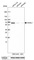
- Experimental details
- Western blot analysis in MCF-7 cells transfected with control siRNA, target specific siRNA probe #1 and #2, using Anti-VANGL1 antibody. Remaining relative intensity is presented. Loading control: Anti-GAPDH.
- Sample type
- Human
- Protocol
- Protocol

- Submitted by
- Atlas Antibodies (provider)
- Enhanced method
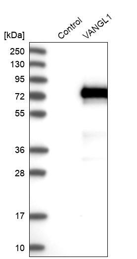
- Recombinant expression validation
- Main image

- Experimental details
- Western blot analysis in control (vector only transfected HEK293T lysate) and VANGL1 over-expression lysate (Co-expressed with a C-terminal myc-DDK tag (~3.1 kDa) in mammalian HEK293T cells, LY403377).
- Sample type
- Human
- Protocol
- Protocol
Supportive validation
- Submitted by
- Atlas Antibodies (provider)
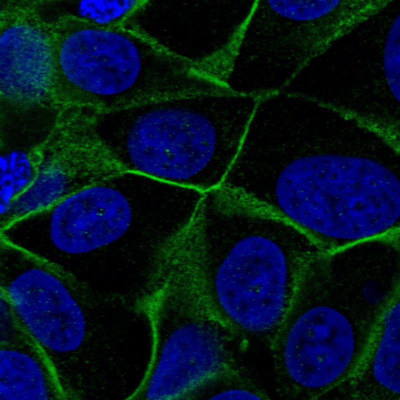
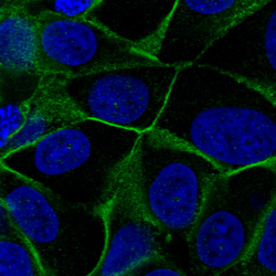
- Main image
- Experimental details
- Immunofluorescent staining of MCF7 cells shows localization to plasma membrane.
- Sample type
- Human