 Explore
 Validate
 Learn
Learn Western blot
 Immunocytochemistry
Antibody data
- Antibody Data
- Antigen structure
- References [1]
- Comments [0]
- Validations
- Immunocytochemistry [2]
- Chromatin Immunoprecipitation [2]
Submit
Validation data
Reference
Comment
Report error
- Product number
- 712465 - Provider product page

- Provider
- Invitrogen Antibodies
- Product name
- HJURP Recombinant Superclonal™ Antibody (16HCLC)
- Antibody type
- Other
- Antigen
- Other
- Description
- This antibody is predicted to react with Monkey, Pig, Cat. Recombinant rabbit Superclonal™ antibodies are unique offerings from Thermo Fisher Scientific. They are comprised of a selection of multiple different recombinant monoclonal antibodies, providing the best of both worlds - the sensitivity of polyclonal antibodies with the specificity of monoclonal antibodies - all delivered with the consistency only found in a recombinant antibody. While functionally the same as a polyclonal antibody - recognizing multiple epitope sites on the target and producing higher detection sensitivity for low abundance targets - a recombinant rabbit Superclonal™ antibody has a known mixture of light and heavy chains. The exact population can be produced in every lot, circumventing the biological variability typically associated with polyclonal antibody production. Note: Formerly called “Recombinant polyclonal antibody”, this product is now rebranded as “Recombinant Superclonal™ antibody”. The physical product and the performance remain unchanged.
- Reactivity
- Human
- Host
- Rabbit
- Isotype
- IgG
- Antibody clone number
- 16HCLC
- Vial size
- 100 μg
- Concentration
- 0.5 mg/mL
- Storage
- Store at 4°C short term. For long term storage, store at -20°C, avoiding freeze/thaw cycles.
Submitted references HJURP regulates cell proliferation and chemo-resistance via YAP1/NDRG1 transcriptional axis in triple-negative breast cancer.
Mao M, Jia Y, Chen Y, Yang J, Xu L, Zhang X, Zhou J, Li Z, Chen C, Ju S, Wang L
Cell death & disease 2022 Apr 22;13(4):396
Cell death & disease 2022 Apr 22;13(4):396
No comments: Submit comment
Supportive validation
- Submitted by
- Invitrogen Antibodies (provider)
- Main image
- Experimental details
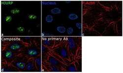
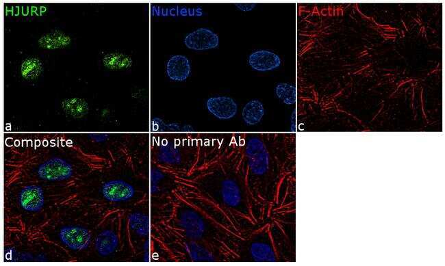
- For immunofluorescence analysis, A549 cells were fixed and permeabilized for detection of endogenous HJURP using Anti-HJURP Recombinant Rabbit Polyclonal Antibody (Product # 712465, 1:100 dilution) and labeled with Goat anti-Rabbit IgG (H+L) Superclonal™ Secondary Antibody, Alexa Fluor® 488 conjugate (Product # A27034, 1:2000). Panel a) shows representative cells that were stained for detection and localization of HJURP protein (green), Panel b) is stained for nuclei (blue) using ProLong™ Diamond Antifade Mountant with DAPI (Product # P36962). Panel c) represents cytoskeletal F-actin staining using Rhodamine Phalloidin (Product # R415, 1:300). Panel d) is a composite image of Panels a, b and c clearly demonstrating nuclear localization of HJURP. Panel e) represents control cells with no primary antibody to assess background. The images were captured at 60X magnification.
- Submitted by
- Invitrogen Antibodies (provider)
- Main image

- Experimental details
- For immunofluorescence analysis, A549 cells were fixed and permeabilized for detection of endogenous HJURP using Anti-HJURP Recombinant Rabbit Superclonal™ Antibody (Product # 712465, 1:100 dilution) and labeled with Goat anti-Rabbit IgG (Heavy Chain) Superclonal™ Secondary Antibody, Alexa Fluor® 488 conjugate (Product # A27034, 1:2000). Panel a) shows representative cells that were stained for detection and localization of HJURP protein (green), Panel b) is stained for nuclei (blue) using ProLong™ Diamond Antifade Mountant with DAPI (Product # P36962). Panel c) represents cytoskeletal F-actin staining using Rhodamine Phalloidin (Product # R415, 1:300). Panel d) is a composite image of Panels a, b and c clearly demonstrating nuclear localization of HJURP. Panel e) represents control cells with no primary antibody to assess background. The images were captured at 60X magnification.

Supportive validation
- Submitted by
- Invitrogen Antibodies (provider)
- Main image

- Experimental details
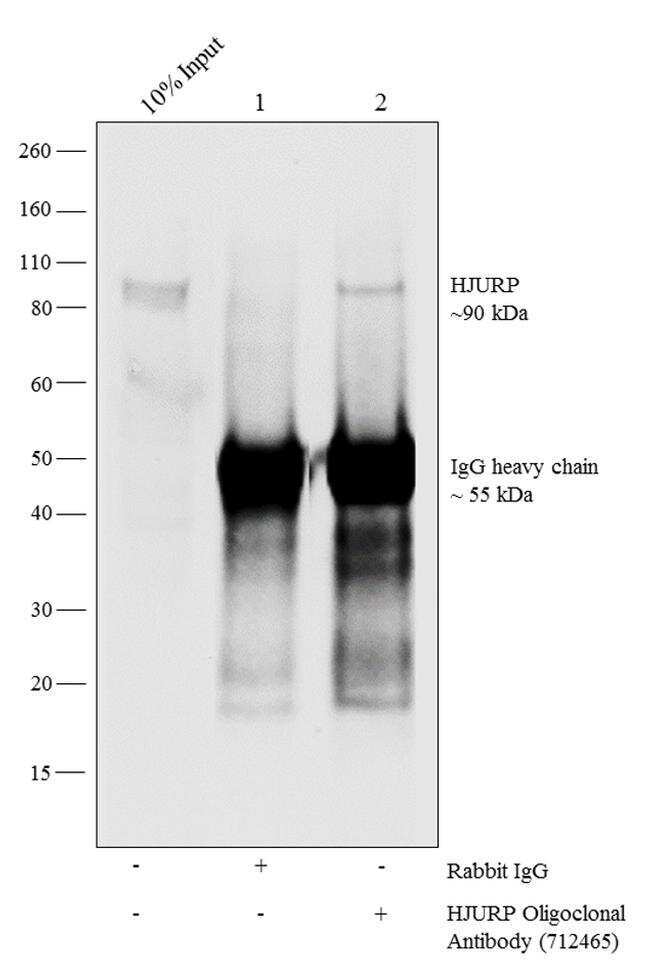
- Chromatin Immunoprecipitation-Western blot (ChIP-WB) of endogenous HJURP protein using Anti-HJURP Antibody: ChIP was performed using Anti-HJURP Recombinant Rabbit Polyclonal Antibody (Product # 712465, 10 µg) (Lane 3) on sheared chromatin from 4 million formaldehyde fixed A549 cells. Normal Rabbit IgG (lane 2) was used as negative IP control. Western blot analysis of immunoprecipitated proteins was performed using Anti-HJURP Recombinant Rabbit Polyclonal Antibody (Product # 712465, 1:200 dilution).
- Submitted by
- Invitrogen Antibodies (provider)
- Main image

- Experimental details
- Chromatin Immunoprecipitation-Western blot (ChIP-WB) of endogenous HJURP protein using Anti-HJURP Antibody: ChIP was performed using Anti-HJURP Recombinant Rabbit Superclonal™ Antibody (Product # 712465, 10 µg) (Lane 3) on sheared chromatin from 4 million formaldehyde fixed A549 cells. Normal Rabbit IgG (lane 2) was used as negative IP control. Western blot analysis of immunoprecipitated proteins was performed using Anti-HJURP Recombinant Rabbit Superclonal™ Antibody (Product # 712465, 1:200 dilution).
