Explore
 Validate
 Learn
LearnMA5800
antibody from Invitrogen Antibodies
Targeting: CD58
LFA3
 Western blot
 Immunocytochemistry
 Immunoprecipitation Immunohistochemistry Flow cytometry Blocking/Neutralizing
Antibody data
- Antibody Data
- Antigen structure
- References [15]
- Comments [0]
- Validations
- Immunocytochemistry [2]
- Flow cytometry [5]
Submit
Validation data
Reference
Comment
Report error
- Product number
- MA5800 - Provider product page

- Provider
- Invitrogen Antibodies
- Product name
- CD58 Monoclonal Antibody (TS2/9)
- Antibody type
- Monoclonal
- Antigen
- Other
- Description
- MA5800 targets CD58 in WB, FACS, IF, IHC(F), IP, and Neu applications and shows reactivity with Human and Mouse samples. The MA5800 immunogen is human Leukocyte Function Antigen-3. MA5800 detects CD58 which has a predicted molecular weight of approximately 24 kDa. This product has been tested for endotoxins by limulus amoebocyte lysate (LAL) assay and contains an endotoxin concentration of less than or equal to 10 endotoxin units per milligram (EU/mg).
- Reactivity
- Human, Mouse
- Host
- Mouse
- Isotype
- IgG
- Antibody clone number
- TS2/9
- Vial size
- 200 μg
- Concentration
- 1.0 mg/mL
- Storage
- -20°C
Submitted references Endoplasmic Reticulum (ER) Reorganization and Intracellular Retention of CD58 Are Functionally Independent Properties of the Human Cytomegalovirus ER-Resident Glycoprotein UL148.
Nurse-like cells promote CLL survival through LFA-3/CD2 interactions.
Identification and purification of classical Hodgkin cells from lymph nodes by flow cytometry and flow cytometric cell sorting.
Lipopolysaccharide signal transduction in oral keratinocytes--involvement of CD59 but not CD14.
ICOS-ligand, expressed on human endothelial cells, costimulates Th1 and Th2 cytokine secretion by memory CD4+ T cells.
Human lung microvascular endothelial cells activate allogeneic T cells through an LFA-3-dependent, but CD86-independent mechanism.
Different roles of the CD2 and LFA-1 T-cell co-receptors for regulating cytotoxic, proliferative, and cytokine responses of human V gamma 9/V delta 2 T cells.
CD4+ T cell and eosinophil adhesion is mediated by specific ICAM-3 ligation and results in eosinophil activation.
CD4+ T cell and eosinophil adhesion is mediated by specific ICAM-3 ligation and results in eosinophil activation.
The glycosylphosphatidylinositol-anchored form and the transmembrane form of CD58 associate with protein kinases.
Molecular interaction between CD58 and CD2 counter-receptors mediates the ability of monocytes to augment T cell activation by IL-12.
AD2, a human molecule involved in the interaction of T cells with epidermal keratinocytes and thymic epithelial cells.
Differential expression of cell adhesion molecules CD54/CD11a and CD58/CD2 by human melanoma cells and functional role in their interaction with cytotoxic cells.
Immunohistologic analysis of the distribution of cell adhesion molecules within the inflammatory synovial microenvironment.
Three distinct antigens associated with human T-lymphocyte-mediated cytolysis: LFA-1, LFA-2, and LFA-3.
Nguyen CC, Domma AJ, Zhang H, Kamil JP
Journal of virology 2020 Feb 14;94(5)
Journal of virology 2020 Feb 14;94(5)
Nurse-like cells promote CLL survival through LFA-3/CD2 interactions.
Boissard F, Tosolini M, Ligat L, Quillet-Mary A, Lopez F, Fournié JJ, Ysebaert L, Poupot M
Oncotarget 2017 Aug 8;8(32):52225-52236
Oncotarget 2017 Aug 8;8(32):52225-52236
Identification and purification of classical Hodgkin cells from lymph nodes by flow cytometry and flow cytometric cell sorting.
Fromm JR, Kussick SJ, Wood BL
American journal of clinical pathology 2006 Nov;126(5):764-80
American journal of clinical pathology 2006 Nov;126(5):764-80
Lipopolysaccharide signal transduction in oral keratinocytes--involvement of CD59 but not CD14.
Yamamoto T, Nakane T, Doi S, Osaki T
Cellular signalling 2003 Sep;15(9):861-9
Cellular signalling 2003 Sep;15(9):861-9
ICOS-ligand, expressed on human endothelial cells, costimulates Th1 and Th2 cytokine secretion by memory CD4+ T cells.
Khayyamian S, Hutloff A, Büchner K, Gräfe M, Henn V, Kroczek RA, Mages HW
Proceedings of the National Academy of Sciences of the United States of America 2002 Apr 30;99(9):6198-203
Proceedings of the National Academy of Sciences of the United States of America 2002 Apr 30;99(9):6198-203
Human lung microvascular endothelial cells activate allogeneic T cells through an LFA-3-dependent, but CD86-independent mechanism.
Hansen AB, Bouchelouche K, Olesen JD
APMIS : acta pathologica, microbiologica, et immunologica Scandinavica 2001 Dec;109(12):849-56
APMIS : acta pathologica, microbiologica, et immunologica Scandinavica 2001 Dec;109(12):849-56
Different roles of the CD2 and LFA-1 T-cell co-receptors for regulating cytotoxic, proliferative, and cytokine responses of human V gamma 9/V delta 2 T cells.
Wang P, Malkovsky M
Molecular medicine (Cambridge, Mass.) 2000 Mar;6(3):196-207
Molecular medicine (Cambridge, Mass.) 2000 Mar;6(3):196-207
CD4+ T cell and eosinophil adhesion is mediated by specific ICAM-3 ligation and results in eosinophil activation.
Douglas IS, Leff AR, Sperling AI
Journal of immunology (Baltimore, Md. : 1950) 2000 Mar 15;164(6):3385-91
Journal of immunology (Baltimore, Md. : 1950) 2000 Mar 15;164(6):3385-91
CD4+ T cell and eosinophil adhesion is mediated by specific ICAM-3 ligation and results in eosinophil activation.
Douglas IS, Leff AR, Sperling AI
Journal of immunology (Baltimore, Md. : 1950) 2000 Mar 15;164(6):3385-91
Journal of immunology (Baltimore, Md. : 1950) 2000 Mar 15;164(6):3385-91
The glycosylphosphatidylinositol-anchored form and the transmembrane form of CD58 associate with protein kinases.
Itzhaky D, Raz N, Hollander N
Journal of immunology (Baltimore, Md. : 1950) 1998 May 1;160(9):4361-6
Journal of immunology (Baltimore, Md. : 1950) 1998 May 1;160(9):4361-6
Molecular interaction between CD58 and CD2 counter-receptors mediates the ability of monocytes to augment T cell activation by IL-12.
Gollob JA, Li J, Kawasaki H, Daley JF, Groves C, Reinherz EL, Ritz J
Journal of immunology (Baltimore, Md. : 1950) 1996 Sep 1;157(5):1886-93
Journal of immunology (Baltimore, Md. : 1950) 1996 Sep 1;157(5):1886-93
AD2, a human molecule involved in the interaction of T cells with epidermal keratinocytes and thymic epithelial cells.
Bruggers CS, Patel DD, Scearce RM, Whichard LP, Haynes BF, Singer KH
Journal of immunology (Baltimore, Md. : 1950) 1995 Mar 1;154(5):2012-22
Journal of immunology (Baltimore, Md. : 1950) 1995 Mar 1;154(5):2012-22
Differential expression of cell adhesion molecules CD54/CD11a and CD58/CD2 by human melanoma cells and functional role in their interaction with cytotoxic cells.
Altomonte M, Gloghini A, Bertola G, Gasparollo A, Carbone A, Ferrone S, Maio M
Cancer research 1993 Jul 15;53(14):3343-8
Cancer research 1993 Jul 15;53(14):3343-8
Immunohistologic analysis of the distribution of cell adhesion molecules within the inflammatory synovial microenvironment.
Hale LP, Martin ME, McCollum DE, Nunley JA, Springer TA, Singer KH, Haynes BF
Arthritis and rheumatism 1989 Jan;32(1):22-30
Arthritis and rheumatism 1989 Jan;32(1):22-30
Three distinct antigens associated with human T-lymphocyte-mediated cytolysis: LFA-1, LFA-2, and LFA-3.
Sanchez-Madrid F, Krensky AM, Ware CF, Robbins E, Strominger JL, Burakoff SJ, Springer TA
Proceedings of the National Academy of Sciences of the United States of America 1982 Dec;79(23):7489-93
Proceedings of the National Academy of Sciences of the United States of America 1982 Dec;79(23):7489-93
No comments: Submit comment
Supportive validation
- Submitted by
- Invitrogen Antibodies (provider)
- Main image

- Experimental details
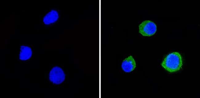
- Immunofluorescent analysis of CD58 (green) showing staining in the cytoplasm of Raji cells (right) compared to a negative control without primary antibody (left). Formalin-fixed cells were permeabilized with 0.1% Triton X-100 in TBS for 5-10 minutes and blocked with 3% BSA-PBS for 30 minutes at room temperature. Cells were probed with a CD58 monoclonal antibody (Product # MA5800) in 3% BSA-PBS at a dilution of 1:20 and incubated overnight at 4ºC in a humidified chamber. Cells were washed with PBST and incubated with a DyLight-conjugated secondary antibody in PBS at room temperature in the dark. F-actin (red) was stained with a fluorescent red phalloidin and nuclei (blue) were stained with Hoechst or DAPI. Images were taken at a magnification of 60x.
- Submitted by
- Invitrogen Antibodies (provider)
- Main image

- Experimental details
- Immunofluorescent analysis of CD58 (green) showing staining in the cytoplasm of Raji cells (right) compared to a negative control without primary antibody (left). Formalin-fixed cells were permeabilized with 0.1% Triton X-100 in TBS for 5-10 minutes and blocked with 3% BSA-PBS for 30 minutes at room temperature. Cells were probed with a CD58 monoclonal antibody (Product # MA5800) in 3% BSA-PBS at a dilution of 1:20 and incubated overnight at 4ºC in a humidified chamber. Cells were washed with PBST and incubated with a DyLight-conjugated secondary antibody in PBS at room temperature in the dark. F-actin (red) was stained with a fluorescent red phalloidin and nuclei (blue) were stained with Hoechst or DAPI. Images were taken at a magnification of 60x.

Supportive validation
- Submitted by
- Invitrogen Antibodies (provider)
- Main image
- Experimental details
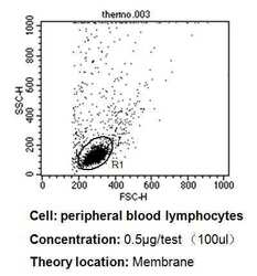
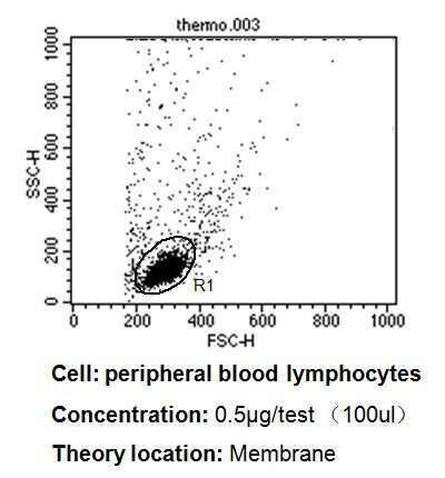
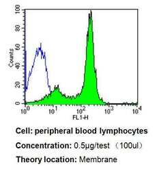
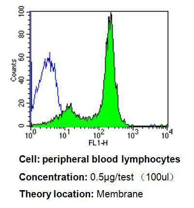
- Flow cytometry analysis of CD58 in PBMC cells compared to an isotype control (blue). Human blood was collected, combined with a hydrophilic polysaccharide, centrifuged, transferred to a conical tube and washed with PBS. 50 µL of cell solution was added to each tube at a dilution of 2x10^7 cells/mL, followed by the addition of 50 µL of isotype control and primary antibody (Product # MA5800) at a dilution of 0.5 µg/test. Cells were incubated for 30 min at 4°C and washed with a cell buffer, followed by incubation with a DyLight 488-conjugated goat anti-mouse IgG (H+L) secondary for 30 min at 4°C in the dark. FACS analysis was performed using 400 µL of cell buffer.
- Submitted by
- Invitrogen Antibodies (provider)
- Main image
- Experimental details
- Flow cytometry analysis of CD58 in PBMC cells compared to an isotype control (blue). Human blood was collected, combined with a hydrophilic polysaccharide, centrifuged, transferred to a conical tube and washed with PBS. 50 µL of cell solution was added to each tube at a dilution of 2x10^7 cells/mL, followed by the addition of 50 µL of isotype control and primary antibody (Product # MA5800) at a dilution of 0.5 µg/test. Cells were incubated for 30 min at 4°C and washed with a cell buffer, followed by incubation with a DyLight 488-conjugated goat anti-mouse IgG (H+L) secondary for 30 min at 4°C in the dark. FACS analysis was performed using 400 µL of cell buffer.
- Submitted by
- Invitrogen Antibodies (provider)
- Main image

- Experimental details
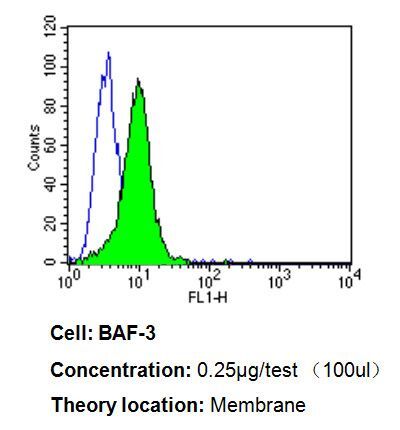
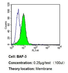
- Flow cytometry analysis of CD58 in BAF-3 cells compared to an isotype control (blue). Cells were harvested, adjusted to a concentration of 1-5x10^6 cells/mL, fixed with 2% paraformaldehyde, washed with PBS, and incubated with CD58 monoclonal antibody (Product # MA5800) at a dilution of 0.25 µg/test for 60 min at room temperature. Cells were then blocked in a solution of 2% BSA-PBS for 30 min at room temperature, incubated for 40 min at room temperature in the dark using a Dylight 488-conjugated goat anti-mouse IgG (H+L) secondary antibody, and re-suspended in PBS for FACS analysis.

- Submitted by
- Invitrogen Antibodies (provider)
- Main image

- Experimental details
- Flow cytometry analysis of CD58 in PBMC cells compared to an isotype control (blue). Human blood was collected, combined with a hydrophilic polysaccharide, centrifuged, transferred to a conical tube and washed with PBS. 50 µL of cell solution was added to each tube at a dilution of 2x10^7 cells/mL, followed by the addition of 50 µL of isotype control and primary antibody (Product # MA5800) at a dilution of 0.5 µg/test. Cells were incubated for 30 min at 4°C and washed with a cell buffer, followed by incubation with a DyLight 488-conjugated goat anti-mouse IgG (H+L) secondary for 30 min at 4°C in the dark. FACS analysis was performed using 400 µL of cell buffer.

- Submitted by
- Invitrogen Antibodies (provider)
- Main image
- Experimental details
- Flow cytometry analysis of CD58 in BAF-3 cells compared to an isotype control (blue). Cells were harvested, adjusted to a concentration of 1-5x10^6 cells/mL, fixed with 2% paraformaldehyde, washed with PBS, and incubated with CD58 monoclonal antibody (Product # MA5800) at a dilution of 0.25 µg/test for 60 min at room temperature. Cells were then blocked in a solution of 2% BSA-PBS for 30 min at room temperature, incubated for 40 min at room temperature in the dark using a Dylight 488-conjugated goat anti-mouse IgG (H+L) secondary antibody, and re-suspended in PBS for FACS analysis.