Explore
 Validate
 Learn
Learn Western blot
Antibody data
- Antibody Data
- Antigen structure
- References [6]
- Comments [0]
- Validations
- Western blot [2]
- Immunocytochemistry [3]
- Immunohistochemistry [2]
- Other assay [7]
Submit
Validation data
Reference
Comment
Report error
- Product number
- PA5-34641 - Provider product page

- Provider
- Invitrogen Antibodies
- Product name
- Periostin Polyclonal Antibody
- Antibody type
- Polyclonal
- Antigen
- Recombinant full-length protein
- Description
- Recommended positive controls: HeLa, A431, H1299, HeLaS3, NIH-3T3. Predicted reactivity: Pig (95%). Store product as a concentrated solution. Centrifuge briefly prior to opening the vial.
- Reactivity
- Human, Mouse
- Host
- Rabbit
- Isotype
- IgG
- Vial size
- 100 μL
- Concentration
- 0.29 mg/mL
- Storage
- Store at 4°C short term. For long term storage, store at -20°C, avoiding freeze/thaw cycles.
Submitted references Single-cell analysis of hepatoblastoma identifies tumor signatures that predict chemotherapy susceptibility using patient-specific tumor spheroids.
Gardenia jasminoides Attenuates Allergic Rhinitis-Induced Inflammation by Inhibiting Periostin Production.
Colocalization of Erythrocytes and Vascular Calcification in Human Atherosclerosis: A Systematic Histomorphometric Analysis.
miR-876 Inhibits EMT and Liver Fibrosis via POSTN to Suppress Metastasis in Hepatocellular Carcinoma.
Periostin Activation of Integrin Receptors on Sensory Neurons Induces Allergic Itch.
NFκB-Induced Periostin Activates Integrin-β3 Signaling to Promote Renal Injury in GN.
Song H, Bucher S, Rosenberg K, Tsui M, Burhan D, Hoffman D, Cho SJ, Rangaswami A, Breese M, Leung S, Ventura MVP, Sweet-Cordero EA, Huang FW, Nijagal A, Wang B
Nature communications 2022 Aug 25;13(1):4878
Nature communications 2022 Aug 25;13(1):4878
Gardenia jasminoides Attenuates Allergic Rhinitis-Induced Inflammation by Inhibiting Periostin Production.
Pyun BJ, Lee JY, Kim YJ, Ji KY, Jung DH, Park KS, Jo K, Choi S, Jung MA, Kim YH, Kim T
Pharmaceuticals (Basel, Switzerland) 2021 Sep 28;14(10)
Pharmaceuticals (Basel, Switzerland) 2021 Sep 28;14(10)
Colocalization of Erythrocytes and Vascular Calcification in Human Atherosclerosis: A Systematic Histomorphometric Analysis.
Böhm EW, Pavlaki M, Chalikias G, Mikroulis D, Georgiadis GS, Tziakas DN, Konstantinides S, Schäfer K
TH open : companion journal to thrombosis and haemostasis 2021 Apr;5(2):e113-e124
TH open : companion journal to thrombosis and haemostasis 2021 Apr;5(2):e113-e124
miR-876 Inhibits EMT and Liver Fibrosis via POSTN to Suppress Metastasis in Hepatocellular Carcinoma.
Chen K, Li Z, Zhang M, Wang B, Peng T, Shen Y, Zhang J, Ye J, Liu Y, Tang D, Peng M, Ma D, Xiao Z, Zhang Y, Jin W, Li X
BioMed research international 2020;2020:1964219
BioMed research international 2020;2020:1964219
Periostin Activation of Integrin Receptors on Sensory Neurons Induces Allergic Itch.
Mishra SK, Wheeler JJ, Pitake S, Ding H, Jiang C, Fukuyama T, Paps JS, Ralph P, Coyne J, Parkington M, DeBrecht J, Ehrhardt-Humbert LC, Cruse GP, Bäumer W, Ji RR, Ko MC, Olivry T
Cell reports 2020 Apr 7;31(1):107472
Cell reports 2020 Apr 7;31(1):107472
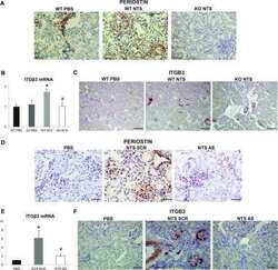
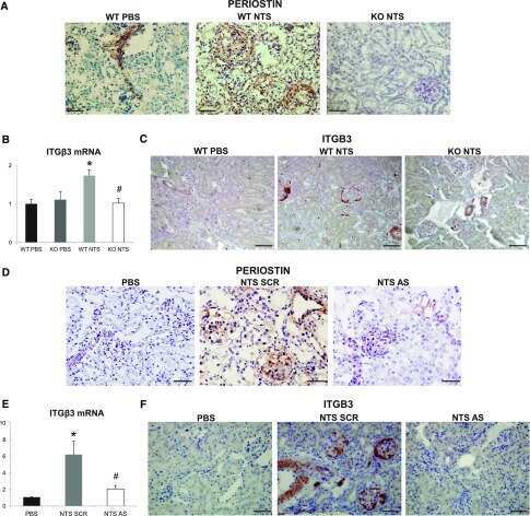
NFκB-Induced Periostin Activates Integrin-β3 Signaling to Promote Renal Injury in GN.
Prakoura N, Kavvadas P, Kormann R, Dussaule JC, Chadjichristos CE, Chatziantoniou C
Journal of the American Society of Nephrology : JASN 2017 May;28(5):1475-1490
Journal of the American Society of Nephrology : JASN 2017 May;28(5):1475-1490
No comments: Submit comment
Supportive validation
- Submitted by
- Invitrogen Antibodies (provider)
- Main image

- Experimental details
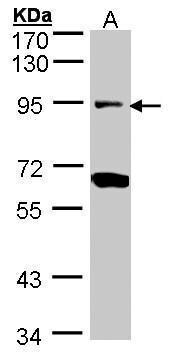
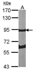
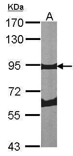
- Western Blot analysis of Periostin was performed by separating 30 µg of H1299 lysates by 7.5% SDS PAGE. Proteins were transferred to a membrane and probed with a Periostin Polyclonal Antibody (Product # PA5-34641) at a dilution of 1:1000. The HRP-conjugated anti-rabbit IgG antibody was used to detect the primary antibody.
- Submitted by
- Invitrogen Antibodies (provider)
- Main image
- Experimental details
- Western Blot analysis of Periostin was performed by separating 30 µg of NIH-3T3 lysates by 7.5% SDS PAGE. Proteins were transferred to a membrane and probed with a Periostin Polyclonal Antibody (Product # PA5-34641) at a dilution of 1:1000. The HRP-conjugated anti-rabbit IgG antibody was used to detect the primary antibody.
Supportive validation
- Submitted by
- Invitrogen Antibodies (provider)
- Main image
- Experimental details
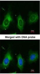
- Immunofluorescent analysis of paraformaldehyde-fixed HeLa cells using anti-Periostin Polyclonal Antibody (Product # PA5-34641) at a 1:200 dilution.
- Submitted by
- Invitrogen Antibodies (provider)
- Main image

- Experimental details
- Immunocytochemistry-Immunofluorescence analysis of Periostin in paraformaldehyde-fixed HeLa cells using Periostin Polyclonal Antibody (Product # PA5-34641) at a dilution of 1:200.
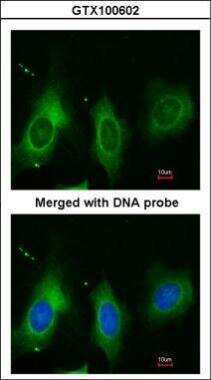
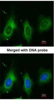
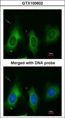
- Submitted by
- Invitrogen Antibodies (provider)
- Main image

- Experimental details
- Immunocytochemistry-Immunofluorescence analysis of Periostin in paraformaldehyde-fixed HeLa cells using Periostin Polyclonal Antibody (Product # PA5-34641) at a dilution of 1:200.
Supportive validation
- Submitted by
- Invitrogen Antibodies (provider)
- Main image
- Experimental details
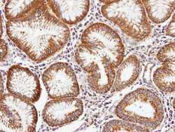
- Immunohistochemistry (Paraffin) analysis of Periostin was performed in paraffin-embedded human gastric tissue using Periostin Polyclonal Antibody (Product # PA5-34641) at a dilution of 1:100. Antigen Retrieval: EDTA based buffer, pH 8.0, 15 min.
- Submitted by
- Invitrogen Antibodies (provider)
- Main image

- Experimental details
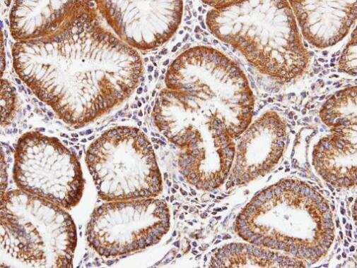
- Immunohistochemistry (Paraffin) analysis of Periostin was performed in paraffin-embedded human gastric tissue using Periostin Polyclonal Antibody (Product # PA5-34641) at a dilution of 1:100. Antigen Retrieval: EDTA based buffer, pH 8.0, 15 min.

Supportive validation
- Submitted by
- Invitrogen Antibodies (provider)
- Main image
- Experimental details
- NULL
- Submitted by
- Invitrogen Antibodies (provider)
- Main image
- Experimental details
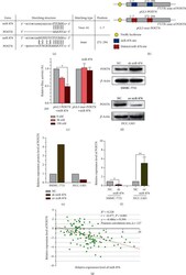
- Figure 3 miR-876 regulates the expression of POSTN. (a) The prediction for miR-876 binding sites on POSTN transcript. (b) Schematic outlining the wild-type and mut-POSTN luciferase plasmid. (c) Luciferase activity in HEK-293 cells cotransfected with indicated miR-876 concentration or indicated POSTN luciferase reporter transcript. Data are showed as the ratio of firefly activity to Renilla luciferase activity. (d, e) The protein level of POSTN was measured by WB assays in indicated treated SMMC-7721 or HCC-LM3 cells. (f) The mRNA expression of POSTN was detected in indicated treated SMMC-7721 or HCC-LM3 cells by qRT-PCR. (g) The correlation analysis of POSTN and miR-876 in 127 tissues of HCC patients.
- Submitted by
- Invitrogen Antibodies (provider)
- Main image
- Experimental details
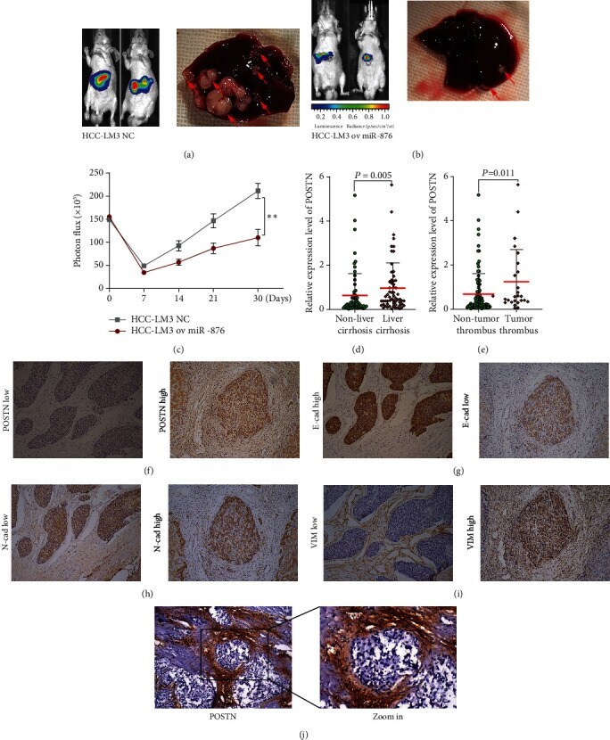
- Figure 5 POSTN was associated with EMT and liver cirrhosis in clinical HCC tissues. (a-c) Animal experiments, the luciferase intensities were measured each week (c) after intracapsular injection with NC (a) or ov miR-876 (b) HCC-LM3 cells in the liver, liver cancer in situ (the red arrows point to), were showed by autopsy (a, b). (d) The expression of POSTN was detected in HCC samples with or without tumor thrombus. (e) The expression of POSTN was detected in HCC samples with or without liver cirrhosis. (f-i) Immunohistochemical staining of POSTN and EMT markers in HCC tissues; representative images of POSTN (f), E-cadherin (g), N-cadherin (h), or vimentin (i) immunostaining of low or high, respectively. (j) Representative image of POSTN immunostaining in HCC with liver cirrhosis tissue.
- Submitted by
- Invitrogen Antibodies (provider)
- Main image
- Experimental details
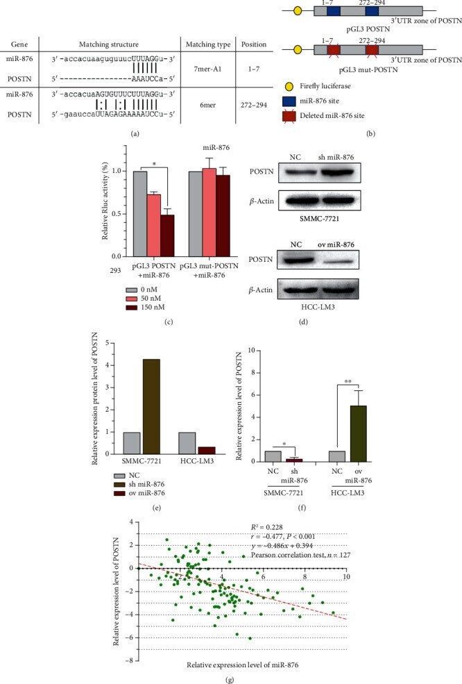
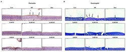
- Figure 3 Effects of Gardenia jasminoides water (GJW) and 70% ethanol (GJE) extracts on histopathological changes in the nasal tissue. Immunohistochemical staining of periostin ( A ) and eosinophil staining ( B ) (upper left corner, scale bar = 50 mum). Con, non-sensitized; AR, allergic rhinitis; DEX, ovalbumin-sensitized with 1 mg/kg dexamethasone.

- Submitted by
- Invitrogen Antibodies (provider)
- Main image
- Experimental details
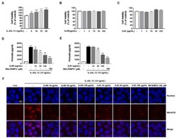
- Figure 5 Effects of Gardenia jasminoides water (GJW) and 70% ethanol (GJE) extracts on interleukin (IL)-4/IL-13-induced periostin generation via mitochondrial reactive oxygen species (ROS) production in human nasal epithelial cells (HNEpCs). Cells were incubated in the presence of the indicated concentrations of IL-4/IL-13, GJW, or GJE for 24 h. ( A - C ) Cell viability was determined using the MTS assay. Cells were treated with GJW or GJE (10, 30, or 100 mug/mL) or MitoTEMPO (100 muM) along with 15 ng/mL IL-4/IL-13 conditioned medium for 24 h ( D , E ) or 1 h ( F ). ( D , E ) Periostin levels in the cell culture medium were measured using a competitive enzyme-linked immunosorbent assay following manufacturer's instructions. ( F ) Analysis of mitochondrial ROS levels in IL-4/IL-13-treated HNEpCs. Cells were incubated with the mitochondrial indicator MitoSOX and analyzed using confocal microscopy (magnification: 130x, scalebar: 20 mum) after stimulation with 15 ng/mL IL-4/IL-13 in the presence or absence of GJW, GJE, or MitoTEMPO (the latter was used as a positive control for inhibiting the effects of IL-4/IL-13). Data are representative of three independent experiments and expressed as the means +- standard deviation; ** p < 0.01 and *** p < 0.001 vs. control; ## p < 0.01; ### p < 0.001 vs. IL-4/IL-13. MtTEMPO, MitoTEMPO.

- Submitted by
- Invitrogen Antibodies (provider)
- Main image
- Experimental details
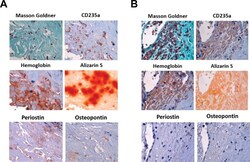
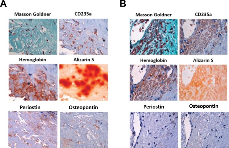
- Fig. 5 Colocalization of lysed and intact erythrocytes and hemoglobin with areas of calcification in serial sections of human vascular lesions. ( A ) Representative example showing the presence of lysed erythrocytes and extracellular hemoglobin in close association with Alizarin S-positive material. ( B ) Representative example showing the presence of intact erythrocytes and cell-associated hemoglobin in an area without calcification; 1,000-fold magnification.
- Submitted by
- Invitrogen Antibodies (provider)
- Main image

- Experimental details
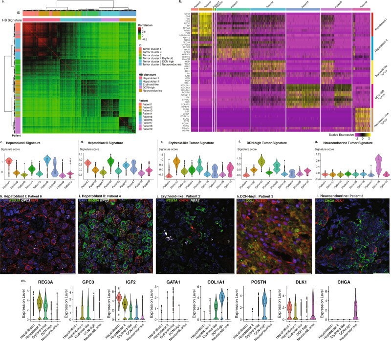
- Tumor cell analysis reveals five transcriptomically distinct tumor signatures detected within the nine HB patients. a Correlation heatmap of the tumor cells by tumor cell clusters, tumor signatures (column annotations), and patients (row annotation). Correlation was shown by the color gradient. Hierarchal clusters were illustrated by dendrograms. b Heatmap of top 10 most differentially expressed genes of each HB signature. Scaled expressed levels are shown by the color bar. c Violin and box plot of the computed c. Hepatoblast I, d Hepatoblast II, e Erythroid-like, f DCN-high, and g Neuroendocrine tumor signature scores for all nine patients ( N = 6244 cells). h FISH staining for REG3A (green), GPC3 (white), and IGF2 (red) of patient 6 tumor tissue (Hepatoblast I), and i patient 4 tumor tissue (Hepatoblast II). j FISH staining for REG3A (green), HBA2 (white), and GATA1 (red), of patient 2 tumor tissue showing Erythroid-like cells (red arrow) and tumor-associated erythroid cells (white arrow). k Immunofluorescence staining for COL1 (green) and POSTN (red) of patient 3 tumor tissue ( DCN -high). l Immunofluorescence staining for CHGA (green) and FISH staining for DLK1 (red) of patient 8 tumor tissue (Neuroendocrine). m Violin plots of individual marker genes presented on panels ( h - l ). Scale bar = 30 um. The box plots present the 25th percentile, the median, the 75th percentile, and outlying or extreme values. The whiskers of the box plots extend to a maximum of 1.5 times the