Explore
 Validate
 Learn
Learn Western blot
Antibody data
- Antibody Data
- Antigen structure
- References [1]
- Comments [0]
- Validations
- Western blot [1]
- Immunoprecipitation [1]
- Immunohistochemistry [1]
Submit
Validation data
Reference
Comment
Report error
- Product number
- AF3548 - Provider product page

- Provider
- R&D Systems
- Product name
- Human Periostin/OSF-2 Antibody
- Antibody type
- Polyclonal
- Description
- Antigen Affinity-purified. Detects human Periostin/OSF-2 in direct ELISAs and Western blots. In direct ELISAs, less than 40% cross-reactivity with recombinant mouse Periostin and recombinant rat Periostin is observed.
- Reactivity
- Human
- Host
- Goat
- Conjugate
- Unconjugated
- Antigen sequence
Q15063- Isotype
- IgG
- Vial size
- 100 ug
- Concentration
- LYOPH
- Storage
- Use a manual defrost freezer and avoid repeated freeze-thaw cycles. 12 months from date of receipt, -20 to -70 °C as supplied. 1 month, 2 to 8 °C under sterile conditions after reconstitution. 6 months, -20 to -70 °C under sterile conditions after reconstitution.
Submitted references Periostin facilitates skin sclerosis via PI3K/Akt dependent mechanism in a mouse model of scleroderma.
Yang L, Serada S, Fujimoto M, Terao M, Kotobuki Y, Kitaba S, Matsui S, Kudo A, Naka T, Murota H, Katayama I
PloS one 2012;7(7):e41994
PloS one 2012;7(7):e41994
No comments: Submit comment
Supportive validation
- Submitted by
- R&D Systems (provider)
- Main image
- Experimental details
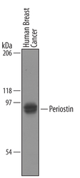
- Detection of Human Periostin/OSF-2 by Western Blot. Western blot shows lysates of human breast cancer tissue. PVDF membrane was probed with 1 µg/mL of Goat Anti-Human Periostin/OSF-2 Antigen Affinity-purified Polyclonal Antibody (Catalog # AF3548) followed by HRP-conjugated Anti-Goat IgG Secondary Antibody (Catalog # HAF019). A specific band was detected for Periostin/OSF-2 at approximately 90-95 kDa (as indicated). This experiment was conducted using Immunoblot Buffer Group 8.

Supportive validation
- Submitted by
- R&D Systems (provider)
- Main image

- Experimental details
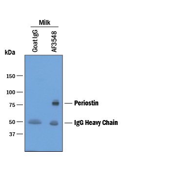
- Immunoprecipitation of Human Periostin/OSF-2. Human Periostin/OSF-2 was immunoprecipitated from human milk samples diluted in 1X Sample Diluent Concentrate 2 (Catalog # DYC002) and incubated with 3 µg Goat Anti-Human Periostin/OSF-2 Antigen Affinity-purified Polyclonal Antibody (Catalog # AF3548) or Normal Goat IgG Control (Catalog # AB-108-C) plus 30 µL Protein G beads overnight. Immunoprecipitated Periostin/OSF-2 was detected by Western blot under reducing conditions using 1 µg/mL Goat Anti-Human Periostin/OSF-2 Antigen Affinity-purified Polyclonal Antibody (Catalog # AF3548). View our recommended buffer recipes for immunoprecipitation.
Supportive validation
- Submitted by
- R&D Systems (provider)
- Main image

- Experimental details
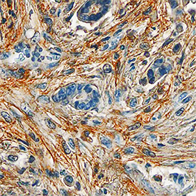
- Periostin/OSF-2 in Human Breast. Periostin/OSF-2 was detected in immersion fixed paraffin-embedded sections of human breast using Goat Anti-Human Periostin/OSF-2 Antigen Affinity-purified Polyclonal Antibody (Catalog # AF3548) at 10 µg/mL overnight at 4 °C. Tissue was stained using the Anti-Goat HRP-DAB Cell & Tissue Staining Kit (brown; Catalog # CTS008) and counterstained with hematoxylin (blue). Specific staining was localized to stromal cells. View our protocol for Chromogenic IHC Staining of Paraffin-embedded Tissue Sections.