 Explore
 Validate
 Learn
Learn Western blot
Antibody data
- Antibody Data
- Antigen structure
- References [2]
- Comments [0]
- Validations
- Western blot [1]
- Immunohistochemistry [1]
Submit
Validation data
Reference
Comment
Report error
- Product number
- PAB10294 - Provider product page

- Provider
- Abnova Corporation
- Proper citation
- Abnova Corporation Cat#PAB10294, RRID:AB_1676309
- Product name
- NOTCH2 polyclonal antibody
- Antibody type
- Polyclonal
- Description
- Rabbit polyclonal antibody raised against synthetic peptide of NOTCH2.
- Storage
- Store at 4°C. For long term storage store at -20°C.Aliquot to avoid repeated freezing and thawing.
Submitted references Notch signaling and inherited disease syndromes.
Notch-1 and Notch-2 exhibit unique patterns of expression in human B-lineage cells.
Gridley T
Human molecular genetics 2003 Apr 1;12 Spec No 1:R9-13
Human molecular genetics 2003 Apr 1;12 Spec No 1:R9-13
Notch-1 and Notch-2 exhibit unique patterns of expression in human B-lineage cells.
Bertrand FE, Eckfeldt CE, Lysholm AS, LeBien TW
Leukemia 2000 Dec;14(12):2095-102
Leukemia 2000 Dec;14(12):2095-102
No comments: Submit comment
Supportive validation
- Submitted by
- Abnova Corporation (provider)
- Main image
- Experimental details
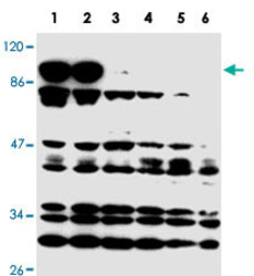
- Western blot using NOTCH2 polyclonal antibody (Cat # PAB10294) (intra) antibody shows detection of a band at ~110 KDa correspondingto active NOTCH2 protein (arrowhead).Western Blot analysis was performed for NOTCH2 expression using 100 ug of total protein lysate obtained from human mesothelial SV40 cells transfected with a plasmid encoding a constitutively active NOTCH2 (intra cellular NOTCH2).Lanes 1-3 contain lysate 24 h (1), 48 h (2), and 72 h (3) post transfection.Lanes 4-6 are the corresponding control cells (untransfected) taken at similar timepoints.The band at about 110 kD represents active NOTCH2.This band is not seen in the control cell.The intracellular domain of NOTCH2 has a predicted band size of 110kD, corresponding to this band.Protein cell lysates were run on a 10% SDS-page gel, blotted onto Hybond C membrane, blocked overnight in PBS-Tween 20 supplemented with 5% Non-fat Milkand probed with NOTCH2 polyclonal antibody at a 1 : 400 dilution.ECL was used as visualization method.

Supportive validation
- Submitted by
- Abnova Corporation (provider)
- Main image
- Experimental details
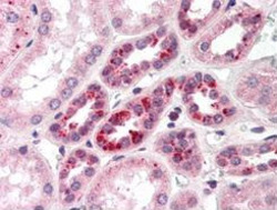
- Immunohistochemical staining with NOTCH2 polyclonal antibody (Cat # PAB10294) was diluted 1 : 500 to detect NOTCH 2 in human kidney tissue. Tissue was formalin fixed and paraffin embedded. No pre-treatment of sample was required. The image shows the localization of antibody as the precipitated red signal, with a hematoxylin purple nuclear counter stain.
- Validation comment
- Immunohistochemistry (Formalin/PFA-fixed paraffin-embedded sections)
