Explore
 Validate
 Learn
Learn Western blot
 Other assay
Antibody data
- Antibody Data
- Antigen structure
- References [1]
- Comments [0]
- Validations
- Other assay [1]
Submit
Validation data
Reference
Comment
Report error
- Product number
- PA5-67829 - Provider product page

- Provider
- Invitrogen Antibodies
- Product name
- ITGA10 Polyclonal Antibody
- Antibody type
- Polyclonal
- Antigen
- Synthetic peptide
- Description
- Predicted to react with Mouse samples.
- Reactivity
- Human, Mouse
- Host
- Rabbit
- Isotype
- IgG
- Vial size
- 100 μL
- Concentration
- 1 mg/mL
- Storage
- -20°C
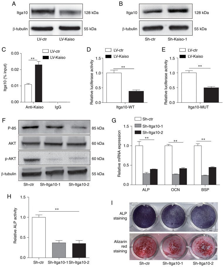
Submitted references Kaiso regulates osteoblast differentiation and mineralization via the Itga10/PI3K/AKT signaling pathway.
Tong W, Li J, Feng X, Wang C, Xu Y, He C, Xu W
International journal of molecular medicine 2021 Apr;47(4)
International journal of molecular medicine 2021 Apr;47(4)
No comments: Submit comment
Supportive validation
- Submitted by
- Invitrogen Antibodies (provider)
- Main image
- Experimental details
- Figure 4 Kaiso regulates the PI3K/AKT pathway via Itga10. Western blot analysis of Itga10 expression in (A) Kaiso-overexpressing or (B) Kaiso-knockdown MC3T3-E1 cells. (C) Chromatin immunoprecipitation analysis of the binding of Kaiso to the promoter of Itga10 in LV-ctr and LV-Kaiso MC3T3-E1 cells. Input DNA and DNA immunoprecipitated with IgG were included as positive and negative controls, respectively. The results are expressed as percentages of the input level. (D) WT and (E) Kaiso binding site-MUT Itga10 promoter-luciferase reporter vectors were co-transfected with Kaiso-overexpression vectors or control vector in MC3T3-E1 cells. Luciferase activity was normalized to Renilla luciferase activity and presented as the fold change to LV-ctr. (F) Expression levels of p85, AKT and p-AKT were determined via western blot analysis in Sh-Itga10-1, Sh-Itga10-2 and Sh-ctr transfectedMC3T3-E1 cells. (G) RT-qPCR analysis of ALP, OCN and BSP expression levels in Sh-ctr and Sh-Kaiso transfected MC3T3-E1 cells after 7 days culture with osteogenic medium. (H) ALP activity was measured on day 7. (I) ALP staining was performed on day 7, and Alizarin Red S staining was performed on day 21. All quantitative data are presented as the mean +- SD (n=3). ** P