 Explore
 Validate
 Learn
Learn Western blot
 Immunohistochemistry
Antibody data
- Antibody Data
- Antigen structure
- References [2]
- Comments [0]
- Validations
- Immunohistochemistry [1]
- Other assay [2]
Submit
Validation data
Reference
Comment
Report error
- Product number
- PA5-20953 - Provider product page

- Provider
- Invitrogen Antibodies
- Product name
- PIAS3 Polyclonal Antibody
- Antibody type
- Polyclonal
- Antigen
- Synthetic peptide
- Description
- A suggested positive control is K562 cell lysate. PA5-20953 can be used with blocking peptide PEP-1067.
- Reactivity
- Human, Mouse, Rat
- Host
- Rabbit
- Isotype
- IgG
- Vial size
- 100 μg
- Concentration
- 1 mg/mL
- Storage
- Maintain refrigerated at 2-8°C for up to 3 months. For long term storage store at -20°C
Submitted references TRIM27 promotes IL-6-induced proliferation and inflammation factor production by activating STAT3 signaling in HaCaT cells.
Protein Inhibitor of Activated STAT3 Suppresses Oxidized LDL-induced Cell Responses during Atherosclerosis in Apolipoprotein E-deficient Mice.
Miao X, Xiang Y, Mao W, Chen Y, Li Q, Fan B
American journal of physiology. Cell physiology 2020 Feb 1;318(2):C272-C281
American journal of physiology. Cell physiology 2020 Feb 1;318(2):C272-C281
Protein Inhibitor of Activated STAT3 Suppresses Oxidized LDL-induced Cell Responses during Atherosclerosis in Apolipoprotein E-deficient Mice.
Wang R, Zhang Y, Xu L, Lin Y, Yang X, Bai L, Chen Y, Zhao S, Fan J, Cheng X, Liu E
Scientific reports 2016 Nov 15;6:36790
Scientific reports 2016 Nov 15;6:36790
No comments: Submit comment
Supportive validation
- Submitted by
- Invitrogen Antibodies (provider)
- Main image
- Experimental details
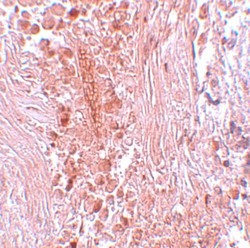
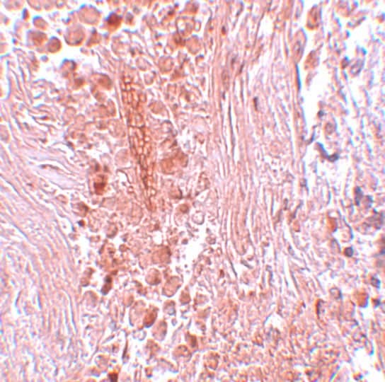
- Immunohistochemistry of PIAS3 in human breast carcinoma tissue with PIAS3 Polyclonal Antibody (Product # PA5-20953) at 5 µg/mL.
Supportive validation
- Submitted by
- Invitrogen Antibodies (provider)
- Main image

- Experimental details
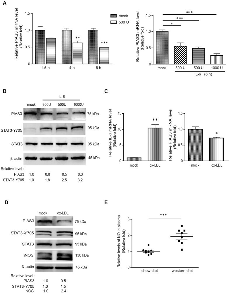
- Figure 2 IL-6 or ox-LDL stimulation reduces PIAS3 expression in RAW264.7 cells. ( A ) IL-6 treatment leads to reductions in PIAS3 expression levels in a time- and dose-dependent manner, as determined via RT-qPCR. ( B ) Detection of PIAS3, STAT3-Y705 and STAT3 protein expression in IL-6-treated cells via Western blotting. Full-length blots were presented in Supplementary Figure S2 . ( C ) Detection of IL-6 and PIAS3 expression in ox-LDL-stimulated cells via RT-qPCR. ( D ) Stimulation with ox-LDL reduces PIAS3 protein expression. PIAS3, STAT3-Y705, STAT3 and iNOS protein expression levels in ox-LDL-treated cells were detected via Western blotting. Full-length blots were presented in Supplementary Figure S3 . ( E ) Relative levels of NO in the plasma of ApoE -/- mice fed a 20-week chow or western diet. Fold changes in NO levels relative to NO levels in mice fed a chow diet are shown (n = 8). Regarding the RT-qPCR results, relative transcript levels are expressed as fold changes compared to transcript levels in mock-treated cells. Regarding the Western blotting results, the relative protein levels in treated cells compared with those in mock-treated cells are shown below the images. *P < 0.05, **P < 0.01, ***P < 0.001.
- Submitted by
- Invitrogen Antibodies (provider)
- Main image
- Experimental details
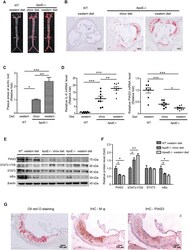
- Figure 1 Atherosclerotic plaque formation is inversely correlated with PIAS3 expression in the aorta. ( A ) Representative images of Oil Red O-stained aortas. ( B ) Representative images showing atherosclerotic plaque formation at the aortic root (bar, 200 mum). ( C ) The relative sizes of the plaque areas at the aortic roots are expressed as fold changes relative to the sizes of the plaque areas of ApoE -/- mice fed a chow diet (n = 8). Serial frozen sections of aortic roots were subjected to Oil Red O staining, and plaque areas were assessed. ( D ) IL-6 and PIAS3 gene expression levels in aortic tissue were determined by RT-qPCR (n = 8). ( E ) PIAS3, STAT3-Y705, STAT3 and IkappaBalpha protein expression levels in aortic tissue were determined by Western blotting. Full-length blots were presented in Supplementary Figure S1 . ( F ) Densitometry analysis of the Western blotting results in panel E. The average protein levels after normalization with beta-actin are expressed as fold changes relative to the protein levels in WT mice fed a western diet. ( G ) Representative images of PIAS3 protein detected in macrophages (Mphi) in atherosclerotic lesions. Serial frozen sections of aortic roots were subjected to immunohistochemical staining with Moma2 (for staining Mphi) and PIAS3 antibodies. Oil Red O staining was also performed to visualize the plaques (bar, 100 mum). *P < 0.05, **P < 0.01, ***P < 0.001.
