Explore
 Validate
 Learn
Learn Western blot
Antibody data
- Antibody Data
- Antigen structure
- References [2]
- Comments [0]
- Validations
- Western blot [2]
- Immunohistochemistry [3]
- Other assay [4]
Submit
Validation data
Reference
Comment
Report error
- Product number
- PA5-93229 - Provider product page

- Provider
- Invitrogen Antibodies
- Product name
- BCL9 Polyclonal Antibody
- Antibody type
- Polyclonal
- Antigen
- Recombinant full-length protein
- Description
- Positive Samples: MCF7, Mouse pancreas; Cellular Location: Nucleus
- Reactivity
- Human, Mouse, Rat
- Host
- Rabbit
- Isotype
- IgG
- Vial size
- 100 µL
- Concentration
- 0.26 mg/mL
- Storage
- -20° C, Avoid Freeze/Thaw Cycles
Submitted references Hypoxia-inducible factor 1α induces osteo/odontoblast differentiation of human dental pulp stem cells via Wnt/β-catenin transcriptional cofactor BCL9.
LncRNA MNX1-AS1 Contributes to Laryngeal Squamous Cell Carcinoma Growth and Migration by Regulating mir-744-5p/bcl9/β-Catenin Axis.
Orikasa S, Kawashima N, Tazawa K, Hashimoto K, Sunada-Nara K, Noda S, Fujii M, Akiyama T, Okiji T
Scientific reports 2022 Jan 13;12(1):682
Scientific reports 2022 Jan 13;12(1):682
LncRNA MNX1-AS1 Contributes to Laryngeal Squamous Cell Carcinoma Growth and Migration by Regulating mir-744-5p/bcl9/β-Catenin Axis.
Ma B, Ren G, Xu J, Yin C, Shi Y
Cell transplantation 2021 Jan-Dec;30:9636897211005682
Cell transplantation 2021 Jan-Dec;30:9636897211005682
No comments: Submit comment
Supportive validation
- Submitted by
- Invitrogen Antibodies (provider)
- Main image

- Experimental details
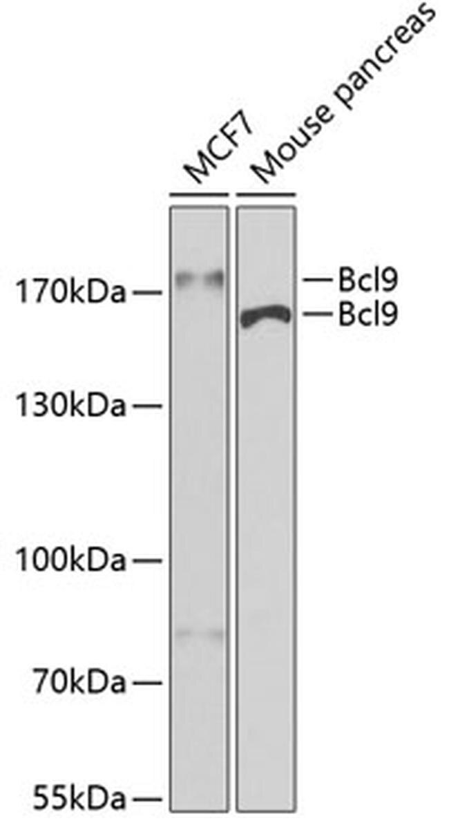
- Western blot analysis of extracts of various cell lines, using Bcl9 Polyclonal antibody (Product # PA5-93229) at 1:400 dilution. Secondary antibody: HRP Goat Anti-Rabbit IgG (H+L) at 1:10000 dilution. Lysates/proteins: 25ug per lane. Blocking buffer: 3% nonfat dry milk in TBST. Exposure time: 30s.
- Submitted by
- Invitrogen Antibodies (provider)
- Main image

- Experimental details
- Western Blot analysis of BCL9 in extracts of various cell lines using BCL9 Polyclonal Antibody (Product # PA5-93229) at a dilution of 1:1000. A HRP Goat Anti-Rabbit IgG (H+L) secondary antibody was used at a dilution of 1:10,000. Lysates/proteins: 25 µg per lane. Blocking buffer: 3% nonfat dry milk in TBST.

Supportive validation
- Submitted by
- Invitrogen Antibodies (provider)
- Main image
- Experimental details
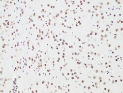
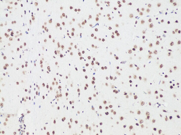
- Immunohistochemistry analysis of BCL9 in paraffin-embedded Rat brain using BCL9 Polyclonal Antibody (Product # PA5-93229) at a dilution of 1:100.
- Submitted by
- Invitrogen Antibodies (provider)
- Main image
- Experimental details
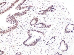
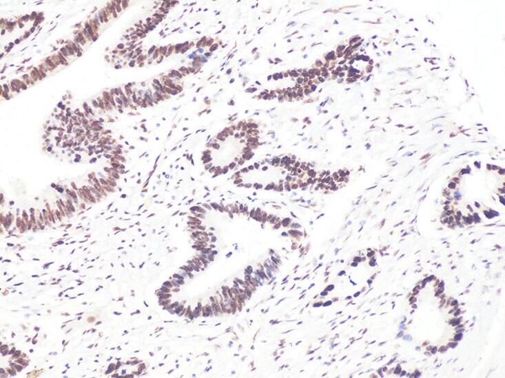
- Immunohistochemistry analysis of BCL9 in paraffin-embedded Human colon carcinoma using BCL9 Polyclonal Antibody (Product # PA5-93229) at a dilution of 1:100.
- Submitted by
- Invitrogen Antibodies (provider)
- Main image
- Experimental details
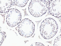
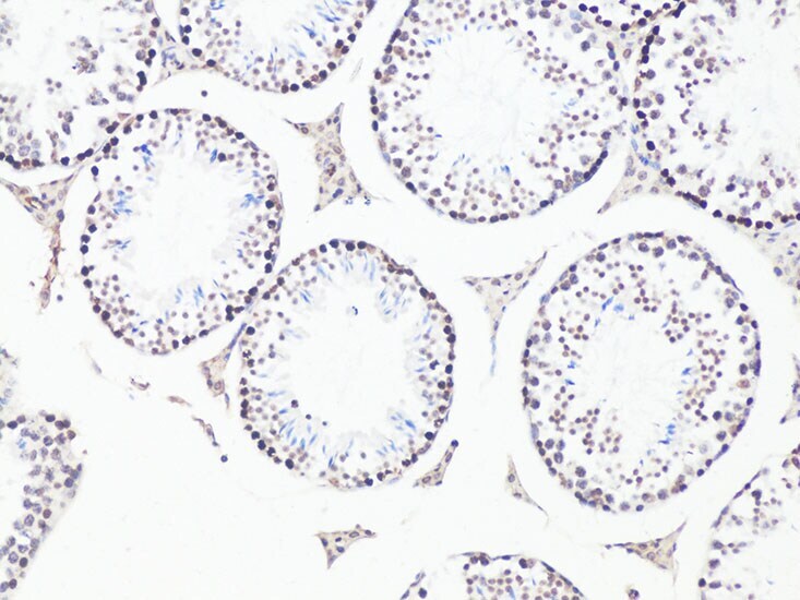
- Immunohistochemistry analysis of BCL9 in paraffin-embedded Mouse testis using BCL9 Polyclonal Antibody (Product # PA5-93229) at a dilution of 1:100.
Supportive validation
- Submitted by
- Invitrogen Antibodies (provider)
- Main image
- Experimental details
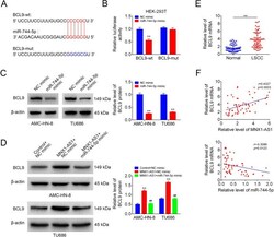
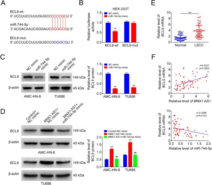
- Figure 4. MNX1-AS1 increased BCL9 expression via targeting miR-744-5p. (A) The putative binding sites between miR-744-5p and BCL9. (B) The luciferase activity of BCL9-wt/mut was detected by luciferase gene reporter Assay ( n = 3, ** P < 0.01, compared with NC mimic group). (C) BCL9 protein levels in NC mimic or miR-744-5p mimic transfected cells were detected by using Western blotting ( n = 3, ** P < 0.01, compared with NC mimic group). (D) BCL9 protein levels in AMC-HN-8 and TU686 cells in control+NC mimic, MNX1-AS1+NC mimic and MNX1-AS1+miR-744-5p mimic groups were detected by Western blotting ( n = 3, ** P < 0.01, compared with control+NC mimic group; ## P < 0.01, compared with mnx1-as1+nc mimic group). (E) QRT-PCR was used to detect the mRNA level of BCL9 in human LSCC tissues and the paired normal tissues ( n = 65, ** P < 0.01, compared with normal group). (F) Correlations between the expression levels of miR-744-5p and BCL9/MNX1-AS1 in 65 cases of human LSCC tissues were determined by Pearson Correlation Analysis.
- Submitted by
- Invitrogen Antibodies (provider)
- Main image
- Experimental details
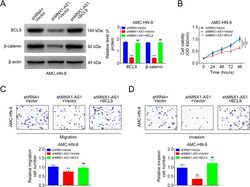
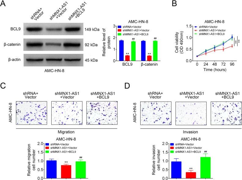
- Figure 5. MNX1-AS1 inhibited LSCC cell viability, migration and invasion via inhibiting BCL9/beta-catenin signaling. AMC-HN-8 cells in shRNA+vector group, shMNX1-AS1+vector group and shMNX1-AS1+BCL9 groups were submitted to the following assays. (A) Western blotting technology used for the detection of BCL9 and beta-catenin expression levels ( n = 3, ** P < 0.01, compared with shRNA+vector group; ## P < 0.01, compared with shMNX1-AS1+vector group). (B) MTT assay for cell viability detection ( n = 3, ** P < 0.01, compared with shRNA+vector group). (C, D) Transwell chambers used for cell migration and invasion detection ( n = 3, ** P < 0.01, compared with shRNA+vector group; ## P < 0.01, compared with shMNX1-AS1+vector group). LSCC: laryngeal squamous cell carcinoma.
- Submitted by
- Invitrogen Antibodies (provider)
- Main image
- Experimental details
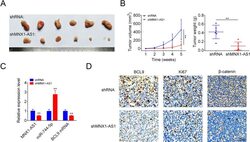
- Figure 6. Knockdown of MNX1-AS1 inhibited cell tumorigenesis in LSCC cells. (A) Tumor shapes in shRNA and shMNX1-AS1 groups. (B) Tumor volumes and weights in shRNA and shMNX1-AS1 groups. (C) The levels of miR-744-5p, MNX1-AS1 and BCL9 mRNA were detected by using qRT-PCR assay in tumor tissues from the shRNA and shMNX1-AS1 groups. (D) Immunohistochemistry technology was used to detect the protein levels of BCL9, Ki-67 and beta-catenin in tumor tissues from the shRNA and shMNX1-AS1 groups. ( ** P < 0.01, compared with shRNA group.). LSCC: laryngeal squamous cell carcinoma.

- Submitted by
- Invitrogen Antibodies (provider)
- Main image

- Experimental details
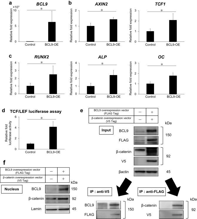
- Figure 4 BCL9 overexpression promotes osteo/odontoblast differentiation and Wnt/beta-catenin signaling. ( a - c ) BCL9 overexpression upregulates the mRNA expression of BCL9, Wnt/beta-catenin target genes (AXIN2 and TCF1), and osteo/odontoblast differentiation markers (RUNX2, ALP, and OC) for 24 h. ( d ) TCF/LEF luciferase assay showing the activation of Wnt/beta-catenin signaling in hDPSCs with BCL9 overexpression for 24 h. ( e ) Immunoprecipitation to detect the binding of BCL9 to beta-catenin in BCL9 (FLAG Tag) and beta-catenin (V5 Tag) overexpression for 24 h. ( f ) BCL9 and beta-catenin protein expression are upregulated in the nucleus of hDPSCs in BCL9 and beta-catenin overexpression for 24 h. Full-length blots are presented in Supplementary Fig. 2 . Error bars indicate standard deviation (n = 4). *p < 0.05.