Explore
 Validate
 Learn
Learn Western blot
 ELISA
ELISAAntibody data
- Antibody Data
- Antigen structure
- References [5]
- Comments [0]
- Validations
- Western blot [1]
- Immunocytochemistry [1]
- Immunohistochemistry [1]
Submit
Validation data
Reference
Comment
Report error
- Product number
- ABIN955341 - Provider product page

- Provider
- antibodies-online
- Product name
- anti-Transient Receptor Potential Cation Channel, Subfamily M, Member 8 (TRPM8) (Center) antibody
- Antibody type
- Polyclonal
- Antigen
- Other
- Reactivity
- Human
- Host
- Rabbit
- Vial size
- 0.1 mg
Submitted references Variation at the NFATC2 locus increases the risk of thiazolidinedione-induced edema in the Diabetes REduction Assessment with ramipril and rosiglitazone Medication (DREAM) study.
Transient receptor potential channel TRPM8 is over-expressed and required for cellular proliferation in pancreatic adenocarcinoma.
Contribution of the S5-pore-S6 domain to the gating characteristics of the cation channels TRPM2 and TRPM8.
TRP channels in human prostate.
PSA reduces prostate cancer cell motility by stimulating TRPM8 activity and plasma membrane expression.
Bailey SD, Xie C, Do R, Montpetit A, Diaz R, Mohan V, Keavney B, Yusuf S, Gerstein HC, Engert JC, Anand S, DREAM investigators
Diabetes care 2010 Oct;33(10):2250-3
Diabetes care 2010 Oct;33(10):2250-3
Transient receptor potential channel TRPM8 is over-expressed and required for cellular proliferation in pancreatic adenocarcinoma.
Yee NS, Zhou W, Lee M
Cancer letters 2010 Nov 1;297(1):49-55
Cancer letters 2010 Nov 1;297(1):49-55
Contribution of the S5-pore-S6 domain to the gating characteristics of the cation channels TRPM2 and TRPM8.
Kühn FJ, Witschas K, Kühn C, Lückhoff A
The Journal of biological chemistry 2010 Aug 27;285(35):26806-14
The Journal of biological chemistry 2010 Aug 27;285(35):26806-14
TRP channels in human prostate.
Van Haute C, De Ridder D, Nilius B
TheScientificWorldJournal 2010 Aug 17;10:1597-611
TheScientificWorldJournal 2010 Aug 17;10:1597-611
PSA reduces prostate cancer cell motility by stimulating TRPM8 activity and plasma membrane expression.
Gkika D, Flourakis M, Lemonnier L, Prevarskaya N
Oncogene 2010 Aug 12;29(32):4611-6
Oncogene 2010 Aug 12;29(32):4611-6
No comments: Submit comment
Supportive validation
- Submitted by
- antibodies-online (provider)
- Main image
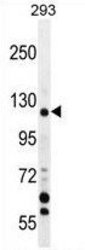
- Experimental details
- TRPM8 Antibody (Center) (AP54364PU-N) western blot analysis in 293 cell line lysates (35 μg/lane). This demonstrates the TRPM8 antibody detected the TRPM8 protein (arrow).

Supportive validation
- Submitted by
- antibodies-online (provider)
- Main image
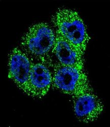
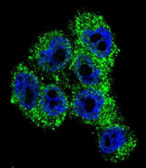
- Experimental details
- Confocal immunofluorescent analysis of TRPM8 Antibody (Center) (AP54364PU-N) with A375 cell followed by Alexa Fluor 488-conjugated goat anti-rabbit lgG (green). DAPI was used to stain the cell nuclear (blue).
Supportive validation
- Submitted by
- antibodies-online (provider)
- Main image

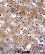
- Experimental details
- TRPM8 Antibody (Center) (AP54364PU-N)immunohistochemistry analysis in formalin fixed and paraffin embedded human liver tissue followed by peroxidase conjugation of the secondary antibody and DAB staining. This data demonstrates the use of TRPM8 Antibody (Center) for immunohistochemistry. Clinical relevance has not been evaluated.