Explore
 Validate
 Learn
Learn Flow cytometry
Antibody data
- Antibody Data
- Antigen structure
- References [4]
- Comments [0]
- Validations
- Flow cytometry [1]
- Other assay [5]
Submit
Validation data
Reference
Comment
Report error
- Product number
- 46-0649-42 - Provider product page

- Provider
- Invitrogen Antibodies
- Product name
- CD64 Monoclonal Antibody (10.1), PerCP-eFluor™ 710, eBioscience™
- Antibody type
- Monoclonal
- Antigen
- Other
- Description
- Description: The 10.1 monoclonal antibody reacts with human CD64 (FcRI), a 75 kDa type I transmembrane protein. CD64 is the high affinity receptor for IgG and is involved in antibody-dependent cell-mediated cytotoxicity (ADCC), phagocytosis, and regulation of cytokine production. Monocytes and macrophages express CD64, while mature granulocytes and lymphocytes are negative. Applications Reported: This 10.1 antibody has been reported for use in flow cytometric analysis. Applications Tested: This 10.1 antibody has been pre-titrated and tested by flow cytometric analysis of normal human peripheral blood cells. This can be used at 5 µL (0.25 µg) per test. A test is defined as the amount (µg) of antibody that will stain a cell sample in a final volume of 100 µL. Cell number should be determined empirically but can range from 10^5 to 10^8 cells/test. PerCP-eFluor® 710 emits at 710 nm and is excited with the blue laser (488 nm); it can be used in place of PerCP-Cyanine5.5. We recommend using a 710/50 bandpass filter, however, the 695/40 bandpass filter is an acceptable alternative. Please make sure that your instrument is capable of detecting this fluorochrome. Fixation: Samples can be stored in IC Fixation Buffer (Product # 00-822-49) (100 µL cell sample + 100 µL IC Fixation Buffer) or 1-step Fix/Lyse Solution (Product # 00-5333-54) for up to 3 days in the dark at 4°C with minimal impact on brightness and FRET efficiency/compensation. Some generalizations regarding fluorophore performance after fixation can be made, but clone specific performance should be determined empirically. Excitation: 488 nm; Emission: 710 nm; Laser: Blue Laser. Filtration: 0.2 µm post-manufacturing filtered.
- Reactivity
- Human
- Host
- Mouse
- Isotype
- IgG
- Antibody clone number
- 10.1
- Vial size
- 100 Tests
- Concentration
- 5 µL/Test
- Storage
- 4° C, store in dark, DO NOT FREEZE!
Submitted references Murlentamab, a Low Fucosylated Anti-Müllerian Hormone Type II Receptor (AMHRII) Antibody, Exhibits Anti-Tumor Activity through Tumor-Associated Macrophage Reprogrammation and T Cell Activation.
Extracorporeal Hemadsorption versus Glucocorticoids during Cardiopulmonary Bypass: A Prospective, Randomized, Controlled Trial.
Patient iPSC-Derived Macrophages to Study Inborn Errors of the IFN-γ Responsive Pathway.
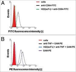
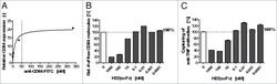
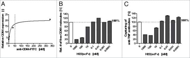
Recombinant H22(scFv) blocks CD64 and prevents the capture of anti-TNF monoclonal antibody. A potential strategy to enhance anti-TNF therapy.
Prat M, Salon M, Allain T, Dubreuil O, Noël G, Preisser L, Jean B, Cassard L, Lemée F, Tabah-Fish I, Pipy B, Jeannin P, Prost JF, Barret JM, Coste A
Cancers 2021 Apr 13;13(8)
Cancers 2021 Apr 13;13(8)
Extracorporeal Hemadsorption versus Glucocorticoids during Cardiopulmonary Bypass: A Prospective, Randomized, Controlled Trial.
Taleska Stupica G, Sostaric M, Bozhinovska M, Rupert L, Bosnic Z, Jerin A, Ihan A, Klokocovnik T, Podbregar M
Cardiovascular therapeutics 2020;2020:7834173
Cardiovascular therapeutics 2020;2020:7834173
Patient iPSC-Derived Macrophages to Study Inborn Errors of the IFN-γ Responsive Pathway.
Haake K, Neehus AL, Buchegger T, Kühnel MP, Blank P, Philipp F, Oleaga-Quintas C, Schulz A, Grimley M, Goethe R, Jonigk D, Kalinke U, Boisson-Dupuis S, Casanova JL, Bustamante J, Lachmann N
Cells 2020 Feb 19;9(2)
Cells 2020 Feb 19;9(2)
Recombinant H22(scFv) blocks CD64 and prevents the capture of anti-TNF monoclonal antibody. A potential strategy to enhance anti-TNF therapy.
Hristodorov D, Mladenov R, Brehm H, Fischer R, Barth S, Thepen T
mAbs 2014;6(5):1283-9
mAbs 2014;6(5):1283-9
No comments: Submit comment
Supportive validation
- Submitted by
- Invitrogen Antibodies (provider)
- Main image

- Experimental details
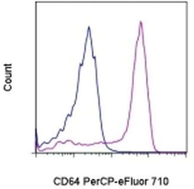
- Staining of normal human peripheral blood cells with Mouse IgG1 K Isotype Control PerCP-eFluor® 710 (Product # 46-4714-82) (blue histogram) or Anti-Human CD64 (Fc gamma Receptor 1) PerCP-eFluor® 710 (purple histogram). Cells in the monocyte gate were used for analysis.
Supportive validation
- Submitted by
- Invitrogen Antibodies (provider)
- Main image
- Experimental details
- NULL

- Submitted by
- Invitrogen Antibodies (provider)
- Main image
- Experimental details
- NULL
- Submitted by
- Invitrogen Antibodies (provider)
- Main image
- Experimental details
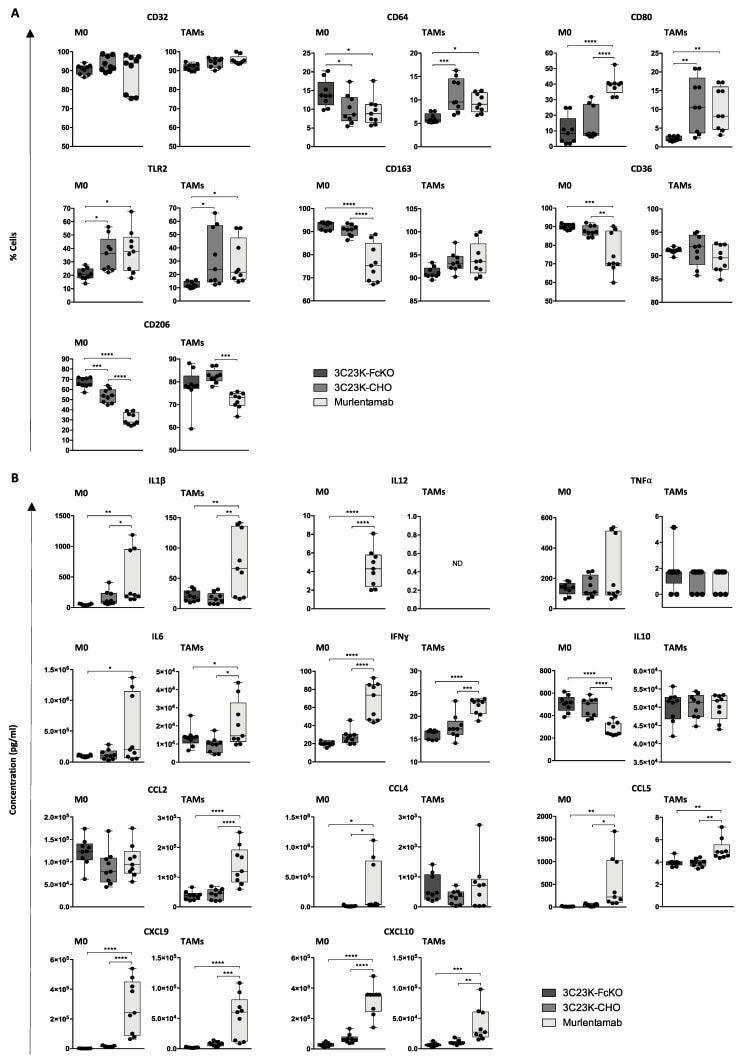
- Figure 3 Murlentamab opsonization of SKOV3-R2 + orients naive macrophages and reprograms TAMs towards an M1-like profile. SKOV3-R2 + ovarian tumor cells were labeled with different 3C23K antibodies (3C23K-FcKO control, 3C23K-CHO normally fucosylated or murlentamab the low fucosylated form) and cultured in the presence of human monocyte-derived macrophages from healthy donors unstimulated (M0) or stimulated with M-CSF and IL-10 (TAMs). ( A ) The proportion of macrophages expressing M1/M2 membrane markers (CD32, CD64, CD80, TLR2, CD163, CD36 and CD206) was determined by flow cytometry after three days of co-culture with SKOV3-R2 + cells. ( B ) The release of cytokines (IL1beta, IL12, TNFalpha, IL6, IFNgamma, IL10) and chemokines (CCL2, CCL4, CCL5, CXCL9 and CXCL10) in the culture medium was determined by AlphaLISA after three days of co-culture with SKOV3-R2 + cells. Data shown (boxplots) are the results from three different experiments (performed with three different healthy donors). * p < 0.05; ** p < 0.01; *** p < 0.001; **** p < 0.0001. p values were determined using one-way ANOVA analysis followed by Tukey''s multiple comparisons test.
- Submitted by
- Invitrogen Antibodies (provider)
- Main image
- Experimental details
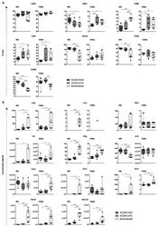
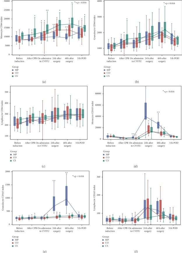
- Figure 3 CD64 and CD163 expression on monocytes, granulocytes, and lymphocytes.
- Submitted by
- Invitrogen Antibodies (provider)
- Main image

- Experimental details
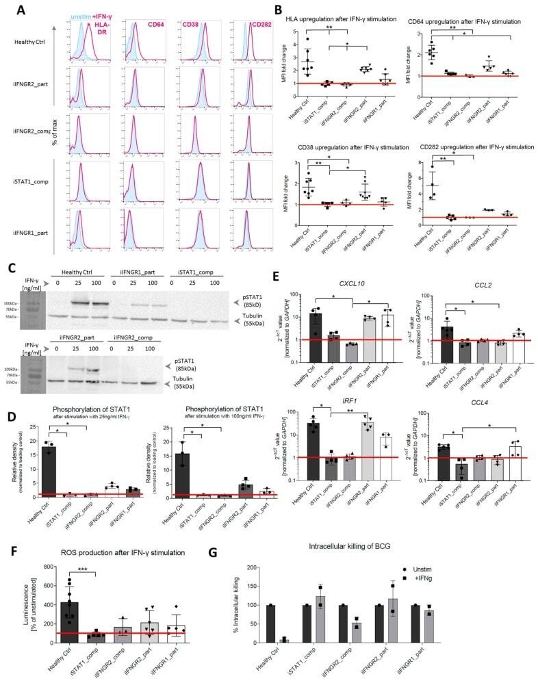
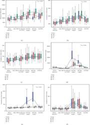
- Figure 4 Patient mutations lead to IFN-gamma-dependent defects in the iPSC-derived macrophages. ( A ) Representative flow cytometric analysis of surface marker (HLA-DR, CD64, CD38, CD282) upregulation after IFN-gamma stimulation in healthy and patient iPSC-derived macrophages. Blue: Isotype. Pink: Surface marker. ( B ) Fold change of median fluorescent intensity (MFI) of HLA-DR, CD64, CD38 and CD282 in healthy and patient iPSC-derived macrophages ( n = 3-7, mean +- SD; each dot represents macrophages from an independent harvest and from at least three independent differentiations, Kruskal-Wallis with Dunn''s multiple comparison)) ( C ) Representative western blot analysis of STAT1 phosphorylation (pSTAT1) after stimulation with low (25 ng/mL) or high (100 ng/mL) dose of IFN-gamma. Tubulin was used as a loading control. The left side of the blot shows the protein size marker. ( D ) Densitometric analysis of STAT1 phosphorylation after IFN-gamma stimulation. Values have been normalized to loading control. ( n = 3, mean +- SD; each dot represents macrophages from an independent harvest and from at least two independent differentiations, Kruskal-Wallis with Dunn''s multiple comparison). ( E ) qRT-PCR analysis of upregulation of downstream targets IRF1, CXCL10, CCL2 and CCL4 after IFNgamma stimulation. Values have been normalized to GAPDH as housekeeping gene. ( n = 3-5, mean +- SD; each dot represents macrophages from an independent harvest and from at least two independent diffe