 Explore
 Validate
 Learn
Learn Western blot
Antibody data
- Antibody Data
- Antigen structure
- References [0]
- Comments [0]
- Validations
- Western blot [2]
- Immunohistochemistry [1]
Submit
Validation data
Reference
Comment
Report error
- Product number
- AF3937 - Provider product page

- Provider
- R&D Systems
- Product name
- Human ECM1 Antibody
- Antibody type
- Polyclonal
- Description
- Antigen Affinity-purified. Detects human ECM-1 in direct ELISAs and Western blots. In direct ELISAs, less than 10% cross-reactivity with recombinant mouse ECM-1 is observed.
- Reactivity
- Human
- Host
- Sheep
- Conjugate
- Unconjugated
- Antigen sequence
AAH23505- Isotype
- IgG
- Vial size
- 100 ug
- Concentration
- LYOPH
- Storage
- Use a manual defrost freezer and avoid repeated freeze-thaw cycles. 12 months from date of receipt, -20 to -70 °C as supplied. 1 month, 2 to 8 °C under sterile conditions after reconstitution. 6 months, -20 to -70 °C under sterile conditions after reconstitution.
No comments: Submit comment
Supportive validation
- Submitted by
- R&D Systems (provider)
- Main image

- Experimental details
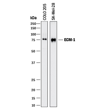
- Detection of Human ECM-1 by Western Blot. Western blot shows lysates of COLO 205 human colorectal adenocarcinoma cell line and SK-Mel-28 human malignant melanoma cell line. PVDF membrane was probed with 1 µg/mL of Sheep Anti-Human ECM-1 Antigen Affinity-purified Polyclonal Antibody (Catalog # AF3937) followed by HRP-conjugated Anti-Sheep IgG Secondary Antibody (Catalog # HAF016). A specific band was detected for ECM-1 at approximately 75 kDa (as indicated). This experiment was conducted under reducing conditions and using Immunoblot Buffer Group 1.
- Submitted by
- R&D Systems (provider)
- Main image

- Experimental details
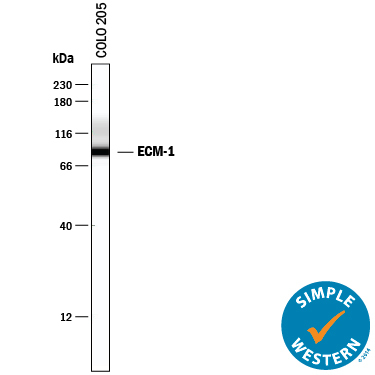
- Detection of Human ECM-1 by Simple WesternTM. Simple Western lane view shows lysates of COLO 205 human colorectal adenocarcinoma cell line, loaded at 0.2 mg/mL. A specific band was detected for ECM-1 at approximately 90 kDa (as indicated) using 10 µg/mL of Sheep Anti-Human ECM-1 Antigen Affinity-purified Polyclonal Antibody (Catalog # AF3937) followed by 1:50 dilution of HRP-conjugated Anti-Sheep IgG Secondary Antibody (Catalog # HAF016). This experiment was conducted under reducing conditions and using the 12-230 kDa separation system.
Supportive validation
- Submitted by
- R&D Systems (provider)
- Main image
- Experimental details
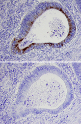
- ECM-1 in Human Colon Cancer Tissue. ECM-1 was detected in immersion fixed paraffin-embedded sections of human colon cancer tissue using 15 µg/mL Sheep Anti-Human ECM-1 Antigen Affinity-purified Polyclonal Antibody (Catalog # AF3937) overnight at 4 °C. Tissue was stained with the Anti-Sheep HRP-DAB Cell & Tissue Staining Kit (brown; Catalog # CTS019) and counterstained with hematoxylin (blue). Specific labeling was localized to the plasma membrane of epithelial cells. Lower panel shows a lack of labeling if primary antibodies are omitted and tissue is stained only with secondary antibody followed by incubation with detection reagents. View our protocol for Chromogenic IHC Staining of Paraffin-embedded Tissue Sections.
