Explore
 Validate
 Learn
LearnF55099-0.4ML
antibody from NSJ Bioreagents
Targeting: GREM1
CKTSF1B1, CRAC1, DAND2, DRM, gremlin, HMPS
 Western blot
Antibody data
- Antibody Data
- Antigen structure
- References [0]
- Comments [0]
- Validations
- Western blot [1]
- Immunohistochemistry [11]
Submit
Validation data
Reference
Comment
Report error
- Product number
- F55099-0.4ML - Provider product page

- Provider
- NSJ Bioreagents
- Product name
- GREM1 Antibody / Gremlin 1
- Antibody type
- Polyclonal
- Description
- Purified
- Reactivity
- Human, Mouse
- Host
- Rabbit
- Vial size
- 0.4 ml, 0.08 ml
- Concentration
- In 1X PBS, pH 7.4, with 0.09% sodium azide
- Storage
- Aliquot the GREM1 antibody and store frozen at -20oC or colder. Avoid repeated freeze-thaw cycles.
No comments: Submit comment
Supportive validation
- Submitted by
- NSJ Bioreagents (provider)
- Main image

- Experimental details
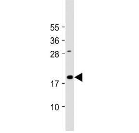
- Western blot testing of mouse skeletal muscle tissue lysate with GREM1 antibody. Predicted molecular weight ~21 kDa.
Supportive validation
- Submitted by
- NSJ Bioreagents (provider)
- Main image
- Experimental details
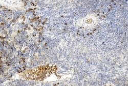
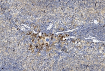
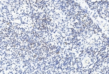
- IHC staining of FFPE mouse spleen tissue with GREM1 antibody. HIER: steam section in pH6 citrate buffer for 20 min and allow to cool prior to staining.

- Submitted by
- NSJ Bioreagents (provider)
- Main image
- Experimental details
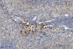
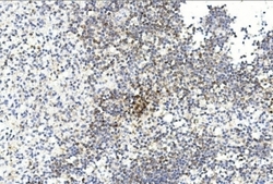
- IHC staining of FFPE mouse spleen tissue with GREM1 antibody. HIER: steam section in pH6 citrate buffer for 20 min and allow to cool prior to staining.
- Submitted by
- NSJ Bioreagents (provider)
- Main image
- Experimental details
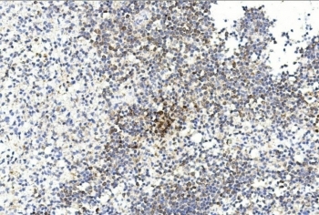
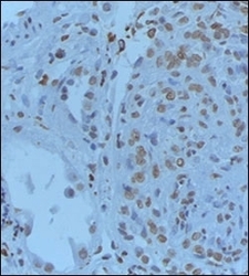
- IHC staining of FFPE human spleen tissue with GREM1 antibody. HIER: steam section in pH6 citrate buffer for 20 min and allow to cool prior to staining.
- Submitted by
- NSJ Bioreagents (provider)
- Main image
- Experimental details
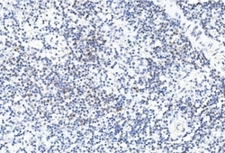
- IHC staining of FFPE human spleen tissue with GREM1 antibody. HIER: steam section in pH6 citrate buffer for 20 min and allow to cool prior to staining.
- Submitted by
- NSJ Bioreagents (provider)
- Main image
- Experimental details
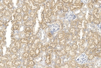
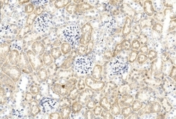
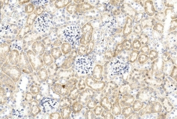
- IHC staining of FFPE human renal tissue with GREM1 antibody. HIER: steam section in pH6 citrate buffer for 20 min and allow to cool prior to staining.

- Submitted by
- NSJ Bioreagents (provider)
- Main image
- Experimental details
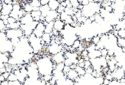
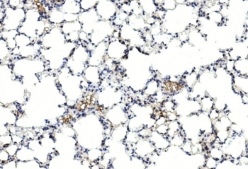
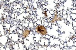
- IHC staining of FFPE mouse lung tissue with GREM1 antibody. HIER: steam section in pH6 citrate buffer for 20 min and allow to cool prior to staining.
- Submitted by
- NSJ Bioreagents (provider)
- Main image
- Experimental details
- IHC staining of FFPE mouse lung tissue with GREM1 antibody. HIER: steam section in pH6 citrate buffer for 20 min and allow to cool prior to staining.

- Submitted by
- NSJ Bioreagents (provider)
- Main image
- Experimental details
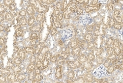
- IHC staining of FFPE mouse kidney tissue with GREM1 antibody. HIER: steam section in pH6 citrate buffer for 20 min and allow to cool prior to staining.
- Submitted by
- NSJ Bioreagents (provider)
- Main image
- Experimental details
- IHC staining of FFPE mouse kidney tissue with GREM1 antibody. HIER: steam section in pH6 citrate buffer for 20 min and allow to cool prior to staining.
- Submitted by
- NSJ Bioreagents (provider)
- Main image
- Experimental details
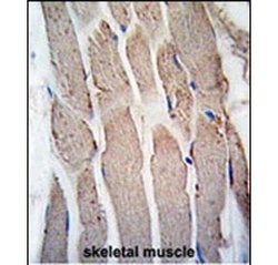
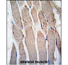
- IHC staining of FFPE human skeletal muscle tissue with GREM1 antibody. HIER: steam section in pH6 citrate buffer for 20 min and allow to cool prior to staining.
- Submitted by
- NSJ Bioreagents (provider)
- Main image
- Experimental details
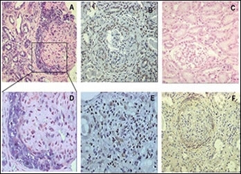
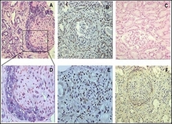
- ISH and IMH demonstrating gremlin mRNA and gremlin protein in crescentic PECs and tubular epithelial cells of patients with pauci-immune crescentic GN. In comparison with normal renal tissue in which there is no expression of gremlin mRNA (C), proliferating PECs of glomerular crescents show a strong expression of gremlin mRNA by ISH (A); and gremlin protein expression by IMH (B); immune competent infiltrating interstitial cells are also strongly positive for gremlin staining (E); and CTGF was also expressed in these glomerular crescentic cells (F).