Explore
 Validate
 Learn
 Flow cytometry
Antibody data
- Antibody Data
- Antigen structure
- References [31]
- Comments [0]
- Validations
- Flow cytometry [2]
- Other assay [18]
Submit
Validation data
Reference
Comment
Report error
- Product number
- 11-9969-42 - Provider product page

- Provider
- Invitrogen Antibodies
- Product name
- CD279 (PD-1) Monoclonal Antibody (MIH4), FITC, eBioscience™
- Antibody type
- Monoclonal
- Antigen
- Other
- Description
- Description: The MIH4 monoclonal antibody reacts with the human PD-1 (programmed death-1), a 55 kDa member of the immunoglobulin superfamily. PD-1 contains the immunoreceptor tyrosine-based inhibitory motif (ITIM) and plays a key role in peripheral tolerance and autoimmune disease. PD-1 is expressed predominantly on activated T and B lymphocytes. Two novel members of the B7 family have been identified as the PD-1 ligands, PD-L1 (B7-H1) and PD-L2 (B7-DC). Evidence reported to date suggests overlapping functions for these two PD-1 ligands and their constitutive expression on some normal tissues and upregulation on activated antigen-presenting cells. The MIH4 antibody recognizes a different epitope than antibody clones J105. Applications Reported: This MIH4 antibody has been reported for use in flow cytometric analysis. Applications Tested: This MIH4 antibody has been pre-titrated and tested by flow cytometric analysis of PHA-stimulated normal human peripheral blood cells. This can be used at 5 µL (2.0 µg) per test. A test is defined as the amount (µg) of antibody that will stain a cell sample in a final volume of 100 µL. Cell number should be determined empirically but can range from 10^5 to 10^8 cells/test. Excitation: 488 nm; Emission: 520 nm; Laser: Blue Laser. Filtration: 0.2 µm post-manufacturing filtered.
- Reactivity
- Human
- Host
- Mouse
- Conjugate
- Green dye
- Isotype
- IgG
- Antibody clone number
- MIH4
- Vial size
- 100 Tests
- Concentration
- 5 μL/Test
- Storage
- 4°C, store in dark, DO NOT FREEZE!
Submitted references Bispecific T-Cell Engaging Antibodies Against MUC16 Demonstrate Efficacy Against Ovarian Cancer in Monotherapy and in Combination With PD-1 and VEGF Inhibition.
Construction of Novel lncRNA-miRNA-mRNA Network Associated With Recurrence and Identification of Immune-Related Potential Regulatory Axis in Hepatocellular Carcinoma.
Imaging Changes and Immune-Checkpoint Expression on T Cells in Bronchoalveolar Lavage Fluid from Patients with Pulmonary Sarcoidosis.
Interaction between HLA-G and NK cell receptor KIR2DL4 orchestrates HER2-positive breast cancer resistance to trastuzumab.
Increased Incidence of Venous Thromboembolism with Cancer Immunotherapy.
PD-1 and BTLA regulate T cell signaling differentially and only partially through SHP1 and SHP2.
Immunogenic neoantigens derived from gene fusions stimulate T cell responses.
Antigen-Presenting Cell-Intrinsic PD-1 Neutralizes PD-L1 in cis to Attenuate PD-1 Signaling in T Cells.
According to Hepatitis C Virus (HCV) Infection Stage, Interleukin-7 Plus 4-1BB Triggering Alone or Combined with PD-1 Blockade Increases TRAF1(low) HCV-Specific CD8(+) Cell Reactivity.
Functional characterization of T-cells from palatine tonsils in patients with chronic tonsillitis.
Structure-guided development of a high-affinity human Programmed Cell Death-1: Implications for tumor immunotherapy.
CXCL13-producing TFH cells link immune suppression and adaptive memory in human breast cancer.
Chronic Critical Illness from Sepsis Is Associated with an Enhanced TCR Response.
Increased levels of CCR7(lo)PD-1(hi) CXCR5(+) CD4(+) T cells, and associated factors Bcl-6, CXCR5, IL-21 and IL-6 contribute to repeated implantation failure.
Tumor-infiltrating Tim-3(+) T cells proliferate avidly except when PD-1 is co-expressed: Evidence for intracellular cross talk.
Immunodominant Dengue Virus-Specific CD8+ T Cell Responses Are Associated with a Memory PD-1+ Phenotype.
IL12-mediated sensitizing of T-cell receptor-dependent and -independent tumor cell killing.
Monoclonal antibodies specific to human Δ42PD1: A novel immunoregulator potentially involved in HIV-1 and tumor pathogenesis.
PD-1(+) CD8(+) T cells are exhausted in tumours and functional in draining lymph nodes of colorectal cancer patients.
IL-17 and IFN-γ expression in lymphocytes from patients with active tuberculosis correlates with the severity of the disease.
Upregulation of programmed death-1 on T cells and programmed death ligand-1 on monocytes in septic shock patients.
The PD-1/PD-L1 axis modulates the natural killer cell versus multiple myeloma effect: a therapeutic target for CT-011, a novel monoclonal anti-PD-1 antibody.
Regulatory T cell (Treg) subsets return in patients with refractory lupus following stem cell transplantation, and TGF-beta-producing CD8+ Treg cells are associated with immunological remission of lupus.
Impaired CD4 and CD8 effector function and decreased memory T cell populations in ICOS-deficient patients.
PD-1/PD-L1 expression in human T-cell leukemia virus type 1 carriers and adult T-cell leukemia/lymphoma patients.
Programmed death-1 expression is associated with the disease status in hepatitis B virus infection.
Programmed death-1 expression is associated with the disease status in hepatitis B virus infection.
Spontaneous recovery in acute human hepatitis C virus infection: functional T-cell thresholds and relative importance of CD4 help.
FOXP3+ Tregs and B7-H1+/PD-1+ T lymphocytes co-infiltrate the tumor tissues of high-risk breast cancer patients: Implication for immunotherapy.
T regulatory cell levels decrease in people infected with Schistosoma mansoni on effective treatment.
PD-1 up-regulation is correlated with HIV-specific memory CD8+ T-cell exhaustion in typical progressors but not in long-term nonprogressors.
Yeku OO, Rao TD, Laster I, Kononenko A, Purdon TJ, Wang P, Cui Z, Liu H, Brentjens RJ, Spriggs D
Frontiers in immunology 2021;12:663379
Frontiers in immunology 2021;12:663379
Construction of Novel lncRNA-miRNA-mRNA Network Associated With Recurrence and Identification of Immune-Related Potential Regulatory Axis in Hepatocellular Carcinoma.
Zhan T, Gao X, Wang G, Li F, Shen J, Lu C, Xu L, Li Y, Zhang J
Frontiers in oncology 2021;11:626663
Frontiers in oncology 2021;11:626663
Imaging Changes and Immune-Checkpoint Expression on T Cells in Bronchoalveolar Lavage Fluid from Patients with Pulmonary Sarcoidosis.
Kotetsu Y, Yanagihara T, Suzuki K, Ando H, Eto D, Hata K, Arimura-Omori M, Yamamoto Y, Harada E, Hamada N
Biomedicines 2021 Sep 16;9(9)
Biomedicines 2021 Sep 16;9(9)
Interaction between HLA-G and NK cell receptor KIR2DL4 orchestrates HER2-positive breast cancer resistance to trastuzumab.
Zheng G, Guo Z, Li W, Xi W, Zuo B, Zhang R, Wen W, Yang AG, Jia L
Signal transduction and targeted therapy 2021 Jun 23;6(1):236
Signal transduction and targeted therapy 2021 Jun 23;6(1):236
Increased Incidence of Venous Thromboembolism with Cancer Immunotherapy.
Roopkumar J, Swaidani S, Kim AS, Thapa B, Gervaso L, Hobbs BP, Wei W, Alban TJ, Funchain P, Kundu S, Sangwan N, Rayman P, Pavicic PG Jr, Diaz-Montero CM, Barnard J, McCrae KR, Khorana AA
Med (New York, N.Y.) 2021 Apr 9;2(4):423-434
Med (New York, N.Y.) 2021 Apr 9;2(4):423-434
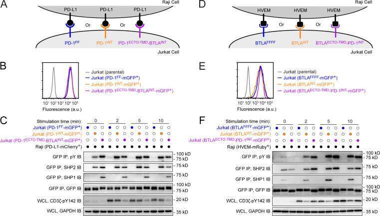
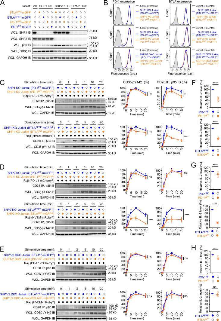
PD-1 and BTLA regulate T cell signaling differentially and only partially through SHP1 and SHP2.
Xu X, Hou B, Fulzele A, Masubuchi T, Zhao Y, Wu Z, Hu Y, Jiang Y, Ma Y, Wang H, Bennett EJ, Fu G, Hui E
The Journal of cell biology 2020 Jun 1;219(6)
The Journal of cell biology 2020 Jun 1;219(6)
Immunogenic neoantigens derived from gene fusions stimulate T cell responses.
Yang W, Lee KW, Srivastava RM, Kuo F, Krishna C, Chowell D, Makarov V, Hoen D, Dalin MG, Wexler L, Ghossein R, Katabi N, Nadeem Z, Cohen MA, Tian SK, Robine N, Arora K, Geiger H, Agius P, Bouvier N, Huberman K, Vanness K, Havel JJ, Sims JS, Samstein RM, Mandal R, Tepe J, Ganly I, Ho AL, Riaz N, Wong RJ, Shukla N, Chan TA, Morris LGT
Nature medicine 2019 May;25(5):767-775
Nature medicine 2019 May;25(5):767-775
Antigen-Presenting Cell-Intrinsic PD-1 Neutralizes PD-L1 in cis to Attenuate PD-1 Signaling in T Cells.
Zhao Y, Harrison DL, Song Y, Ji J, Huang J, Hui E
Cell reports 2018 Jul 10;24(2):379-390.e6
Cell reports 2018 Jul 10;24(2):379-390.e6
According to Hepatitis C Virus (HCV) Infection Stage, Interleukin-7 Plus 4-1BB Triggering Alone or Combined with PD-1 Blockade Increases TRAF1(low) HCV-Specific CD8(+) Cell Reactivity.
Moreno-Cubero E, Subirá D, Sanz-de-Villalobos E, Parra-Cid T, Madejón A, Miquel J, Olveira A, González-Praetorius A, García-Samaniego J, Larrubia JR
Journal of virology 2018 Jan 15;92(2)
Journal of virology 2018 Jan 15;92(2)
Functional characterization of T-cells from palatine tonsils in patients with chronic tonsillitis.
Geißler K, Markwart R, Requardt RP, Weigel C, Schubert K, Scherag A, Rubio I, Guntinas-Lichius O
PloS one 2017;12(9):e0183214
PloS one 2017;12(9):e0183214
Structure-guided development of a high-affinity human Programmed Cell Death-1: Implications for tumor immunotherapy.
Lázár-Molnár E, Scandiuzzi L, Basu I, Quinn T, Sylvestre E, Palmieri E, Ramagopal UA, Nathenson SG, Guha C, Almo SC
EBioMedicine 2017 Mar;17:30-44
EBioMedicine 2017 Mar;17:30-44
CXCL13-producing TFH cells link immune suppression and adaptive memory in human breast cancer.
Gu-Trantien C, Migliori E, Buisseret L, de Wind A, Brohée S, Garaud S, Noël G, Dang Chi VL, Lodewyckx JN, Naveaux C, Duvillier H, Goriely S, Larsimont D, Willard-Gallo K
JCI insight 2017 Jun 2;2(11)
JCI insight 2017 Jun 2;2(11)
Chronic Critical Illness from Sepsis Is Associated with an Enhanced TCR Response.
Borken F, Markwart R, Requardt RP, Schubert K, Spacek M, Verner M, Rückriem S, Scherag A, Oehmichen F, Brunkhorst FM, Rubio I
Journal of immunology (Baltimore, Md. : 1950) 2017 Jun 15;198(12):4781-4791
Journal of immunology (Baltimore, Md. : 1950) 2017 Jun 15;198(12):4781-4791
Increased levels of CCR7(lo)PD-1(hi) CXCR5(+) CD4(+) T cells, and associated factors Bcl-6, CXCR5, IL-21 and IL-6 contribute to repeated implantation failure.
Gong Q, Zhu Y, Pang N, Ai H, Gong X, La X, Ding J
Experimental and therapeutic medicine 2017 Dec;14(6):5931-5941
Experimental and therapeutic medicine 2017 Dec;14(6):5931-5941
Tumor-infiltrating Tim-3(+) T cells proliferate avidly except when PD-1 is co-expressed: Evidence for intracellular cross talk.
Li J, Shayan G, Avery L, Jie HB, Gildener-Leapman N, Schmitt N, Lu BF, Kane LP, Ferris RL
Oncoimmunology 2016;5(10):e1200778
Oncoimmunology 2016;5(10):e1200778
Immunodominant Dengue Virus-Specific CD8+ T Cell Responses Are Associated with a Memory PD-1+ Phenotype.
de Alwis R, Bangs DJ, Angelo MA, Cerpas C, Fernando A, Sidney J, Peters B, Gresh L, Balmaseda A, de Silva AD, Harris E, Sette A, Weiskopf D
Journal of virology 2016 May;90(9):4771-4779
Journal of virology 2016 May;90(9):4771-4779
IL12-mediated sensitizing of T-cell receptor-dependent and -independent tumor cell killing.
Braun M, Ress ML, Yoo YE, Scholz CJ, Eyrich M, Schlegel PG, Wölfl M
Oncoimmunology 2016 Jul;5(7):e1188245
Oncoimmunology 2016 Jul;5(7):e1188245
Monoclonal antibodies specific to human Δ42PD1: A novel immunoregulator potentially involved in HIV-1 and tumor pathogenesis.
Cheng L, Tang X, Liu L, Peng J, Nishiura K, Cheung AK, Guo J, Wu X, Tang HY, An M, Zhou J, Cheung KW, Wang H, Guan X, Wu Z, Chen Z
mAbs 2015;7(3):620-9
mAbs 2015;7(3):620-9
PD-1(+) CD8(+) T cells are exhausted in tumours and functional in draining lymph nodes of colorectal cancer patients.
Wu X, Zhang H, Xing Q, Cui J, Li J, Li Y, Tan Y, Wang S
British journal of cancer 2014 Sep 23;111(7):1391-9
British journal of cancer 2014 Sep 23;111(7):1391-9
IL-17 and IFN-γ expression in lymphocytes from patients with active tuberculosis correlates with the severity of the disease.
Jurado JO, Pasquinelli V, Alvarez IB, Peña D, Rovetta AI, Tateosian NL, Romeo HE, Musella RM, Palmero D, Chuluyán HE, García VE
Journal of leukocyte biology 2012 Jun;91(6):991-1002
Journal of leukocyte biology 2012 Jun;91(6):991-1002
Upregulation of programmed death-1 on T cells and programmed death ligand-1 on monocytes in septic shock patients.
Zhang Y, Li J, Lou J, Zhou Y, Bo L, Zhu J, Zhu K, Wan X, Cai Z, Deng X
Critical care (London, England) 2011;15(1):R70
Critical care (London, England) 2011;15(1):R70
The PD-1/PD-L1 axis modulates the natural killer cell versus multiple myeloma effect: a therapeutic target for CT-011, a novel monoclonal anti-PD-1 antibody.
Benson DM Jr, Bakan CE, Mishra A, Hofmeister CC, Efebera Y, Becknell B, Baiocchi RA, Zhang J, Yu J, Smith MK, Greenfield CN, Porcu P, Devine SM, Rotem-Yehudar R, Lozanski G, Byrd JC, Caligiuri MA
Blood 2010 Sep 30;116(13):2286-94
Blood 2010 Sep 30;116(13):2286-94
Regulatory T cell (Treg) subsets return in patients with refractory lupus following stem cell transplantation, and TGF-beta-producing CD8+ Treg cells are associated with immunological remission of lupus.
Zhang L, Bertucci AM, Ramsey-Goldman R, Burt RK, Datta SK
Journal of immunology (Baltimore, Md. : 1950) 2009 Nov 15;183(10):6346-58
Journal of immunology (Baltimore, Md. : 1950) 2009 Nov 15;183(10):6346-58
Impaired CD4 and CD8 effector function and decreased memory T cell populations in ICOS-deficient patients.
Takahashi N, Matsumoto K, Saito H, Nanki T, Miyasaka N, Kobata T, Azuma M, Lee SK, Mizutani S, Morio T
Journal of immunology (Baltimore, Md. : 1950) 2009 May 1;182(9):5515-27
Journal of immunology (Baltimore, Md. : 1950) 2009 May 1;182(9):5515-27
PD-1/PD-L1 expression in human T-cell leukemia virus type 1 carriers and adult T-cell leukemia/lymphoma patients.
Kozako T, Yoshimitsu M, Fujiwara H, Masamoto I, Horai S, White Y, Akimoto M, Suzuki S, Matsushita K, Uozumi K, Tei C, Arima N
Leukemia 2009 Feb;23(2):375-82
Leukemia 2009 Feb;23(2):375-82
Programmed death-1 expression is associated with the disease status in hepatitis B virus infection.
Ye P, Weng ZH, Zhang SL, Zhang JA, Zhao L, Dong JH, Jie SH, Pang R, Wei RH
World journal of gastroenterology 2008 Jul 28;14(28):4551-7
World journal of gastroenterology 2008 Jul 28;14(28):4551-7
Programmed death-1 expression is associated with the disease status in hepatitis B virus infection.
Ye P, Weng ZH, Zhang SL, Zhang JA, Zhao L, Dong JH, Jie SH, Pang R, Wei RH
World journal of gastroenterology 2008 Jul 28;14(28):4551-7
World journal of gastroenterology 2008 Jul 28;14(28):4551-7
Spontaneous recovery in acute human hepatitis C virus infection: functional T-cell thresholds and relative importance of CD4 help.
Smyk-Pearson S, Tester IA, Klarquist J, Palmer BE, Pawlotsky JM, Golden-Mason L, Rosen HR
Journal of virology 2008 Feb;82(4):1827-37
Journal of virology 2008 Feb;82(4):1827-37
FOXP3+ Tregs and B7-H1+/PD-1+ T lymphocytes co-infiltrate the tumor tissues of high-risk breast cancer patients: Implication for immunotherapy.
Ghebeh H, Barhoush E, Tulbah A, Elkum N, Al-Tweigeri T, Dermime S
BMC cancer 2008 Feb 23;8:57
BMC cancer 2008 Feb 23;8:57
T regulatory cell levels decrease in people infected with Schistosoma mansoni on effective treatment.
Watanabe K, Mwinzi PN, Black CL, Muok EM, Karanja DM, Secor WE, Colley DG
The American journal of tropical medicine and hygiene 2007 Oct;77(4):676-82
The American journal of tropical medicine and hygiene 2007 Oct;77(4):676-82
PD-1 up-regulation is correlated with HIV-specific memory CD8+ T-cell exhaustion in typical progressors but not in long-term nonprogressors.
Zhang JY, Zhang Z, Wang X, Fu JL, Yao J, Jiao Y, Chen L, Zhang H, Wei J, Jin L, Shi M, Gao GF, Wu H, Wang FS
Blood 2007 Jun 1;109(11):4671-8
Blood 2007 Jun 1;109(11):4671-8
No comments: Submit comment
Supportive validation
- Submitted by
- Invitrogen Antibodies (provider)
- Main image
- Experimental details
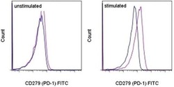
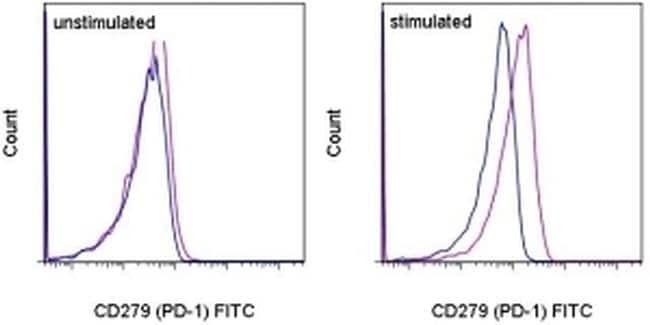
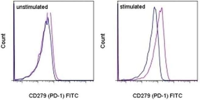
- Staining of unstimulated (left) or 3-day PHA-stimulated (right) normal human peripheral blood cells with Mouse IgG1 K Isotype Control FITC (Product # 11-4714-42) (blue histogram) or Anti-Human CD279 (PD-1) FITC (purple histogram). Total viable cells were used for analysis.
- Conjugate
- Green dye
- Submitted by
- Invitrogen Antibodies (provider)
- Main image
- Experimental details
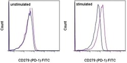
- Staining of unstimulated (left) or 3-day PHA-stimulated (right) normal human peripheral blood cells with Mouse IgG1 K Isotype Control FITC (Product # 11-4714-42) (blue histogram) or Anti-Human CD279 (PD-1) FITC (purple histogram). Total viable cells were used for analysis.
Supportive validation
- Submitted by
- Invitrogen Antibodies (provider)
- Main image
- Experimental details
- NULL
- Conjugate
- Green dye
- Submitted by
- Invitrogen Antibodies (provider)
- Main image
- Experimental details
- NULL
- Conjugate
- Green dye
- Submitted by
- Invitrogen Antibodies (provider)
- Main image
- Experimental details
- NULL
- Conjugate
- Green dye
- Submitted by
- Invitrogen Antibodies (provider)
- Main image
- Experimental details
- NULL
- Conjugate
- Green dye
- Submitted by
- Invitrogen Antibodies (provider)
- Main image
- Experimental details
- NULL
- Conjugate
- Green dye
- Submitted by
- Invitrogen Antibodies (provider)
- Main image
- Experimental details
- NULL
- Conjugate
- Green dye
- Submitted by
- Invitrogen Antibodies (provider)
- Main image
- Experimental details
- NULL
- Conjugate
- Green dye
- Submitted by
- Invitrogen Antibodies (provider)
- Main image
- Experimental details
- NULL
- Conjugate
- Green dye
- Submitted by
- Invitrogen Antibodies (provider)
- Main image
- Experimental details
- NULL
- Conjugate
- Green dye
- Submitted by
- Invitrogen Antibodies (provider)
- Main image
- Experimental details
- NULL
- Conjugate
- Green dye
- Submitted by
- Invitrogen Antibodies (provider)
- Main image
- Experimental details
- NULL
- Conjugate
- Green dye
- Submitted by
- Invitrogen Antibodies (provider)
- Main image
- Experimental details
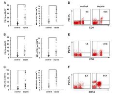
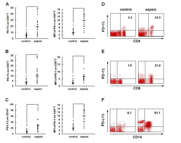
- Figure 2 PD-1 and PD-L1 were upregulated on T cells and monocytes in septic shock patients . Blood samples were obtained from 19 septic shock patients and 22 healthy controls and were stained for programmed death-1 (PD-1) and programmed death ligand-1 (PD-L1) gated on CD4 + T cells, CD8 + T cells, and CD14 + monocytes. (a) to (c) Percentage of PD-1 expression on (a) CD4 + T cells and (b) CD8 + T cells, and (c) percentage of PD-L1 expression on CD14 + monocytes. Each dot represents one individual. Data are mean +- standard error of the mean (SEM) of three independent experiments. ** P < 0.01 compared with healthy controls. (d) to (f) Mean fluorescence intensity (relative fluorescence units) of PD-1 expression on (d) CD4 + T cells, (e) PD-1 expression on CD8 + T cells, and (f) PD-L1 expression on CD14 + monocytes Each dot represents one individual. Data are mean +- SEM of three independent experiments. * P < 0.05 compared with healthy controls. (g) Representative PD-1 expression levels on CD4 + T cells and CD8 + T cells, and PD-L1 expression on CD14 + monocytes. Values in the upper-right quadrant indicate the percentage of cells that express PD-1 or PD-L1.
- Conjugate
- Green dye
- Submitted by
- Invitrogen Antibodies (provider)
- Main image
- Experimental details
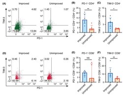
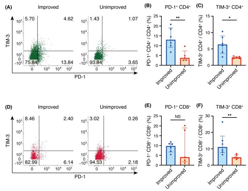
- Figure 2 Programmed cell death 1 (PD-1) and T-cell immunoglobulin- and mucin-domain-containing molecule-3 (TIM-3) expression on CD4 + and CD8 + T cells in bronchoalveolar lavage fluid (BALF). ( A , D ) Representative flow cytometric analysis of PD-1 and TIM-3 expression on CD4 + T cells ( A ) and CD8 + T cells ( D ) in BALF. ( B , E ) Percentages of PD-1 + cells among CD4 + T cells ( B ) and CD8 + T cells ( E ). ( C , F ) Percentages of TIM-3 + cells among CD4 + T cells ( C ) and CD8 + T cells ( F ). ( B , C , E , F ) Individual patient data as well as medians and interquartile ranges are shown. * p < 0.05; ** p < 0.01 (Mann-Whitney U test).
- Conjugate
- Green dye
- Submitted by
- Invitrogen Antibodies (provider)
- Main image
- Experimental details
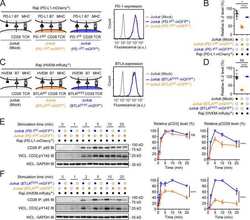
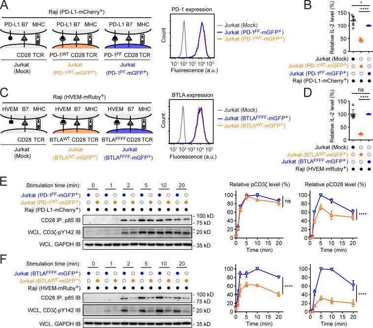
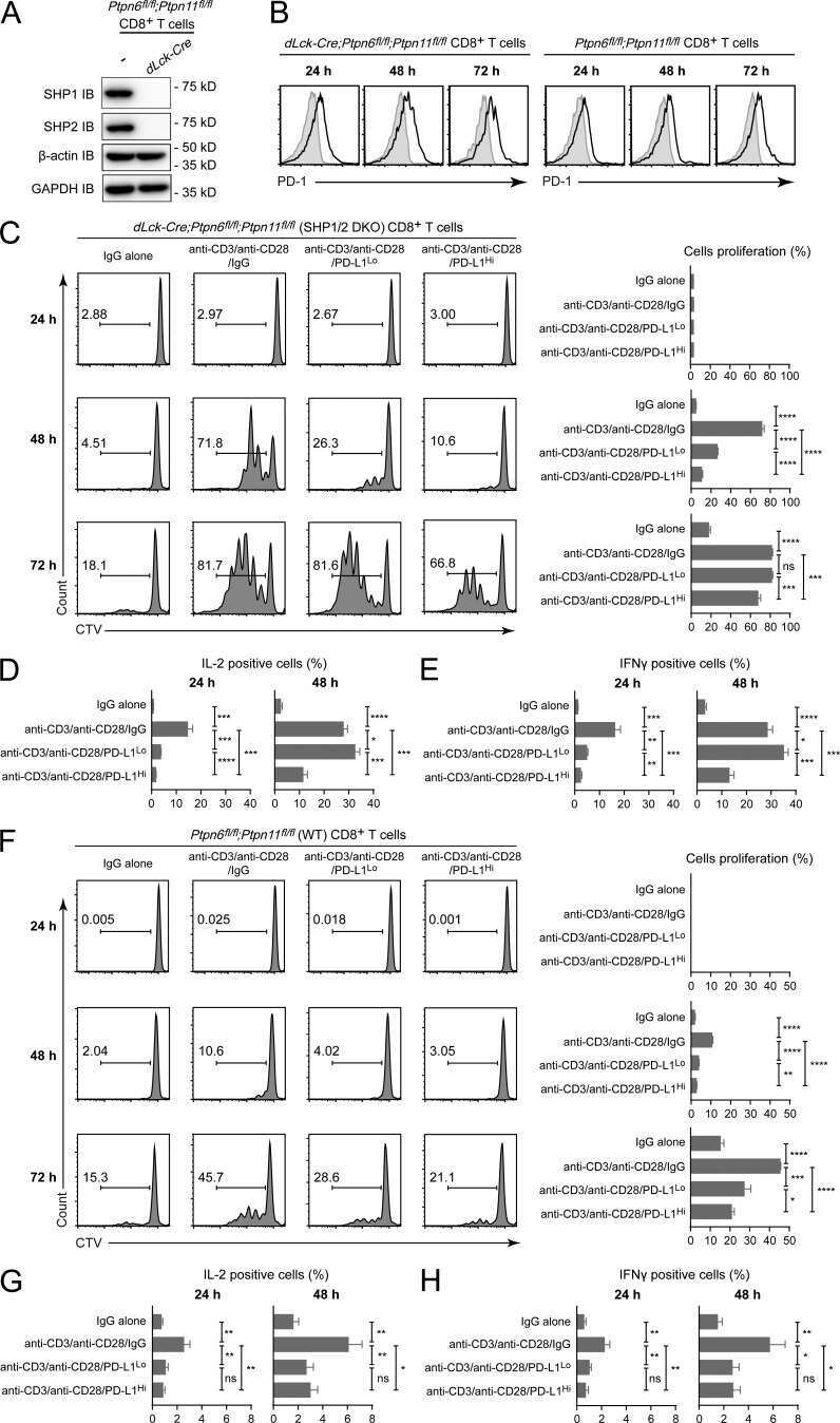
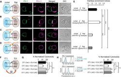
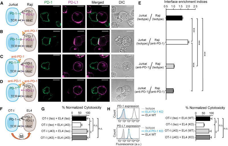
- Figure 7 Blockade of APC-Intrinsic PD-1 Enhances the Synaptic Enrichment of T Cell-Intrinsic PD-1 and Inhibits T Cell-Mediated Cytotoxicity (A) Left: cartoon showing a Jurkat cell expressing PD-1-mGFP (shown as PD-1 in green) conjugated with a Raji cell co-transduced with PD-L1-mCherry and unlabeled PD-1. Right: representative confocal images of the conjugate at the indicated channel acquired 2 min after cell-cell contact. Scale bars, 10 um. (B-D) Same as (A), except that Raji (PD-L1+/PD-1+) cells (B), Jurkat (PD-1-mGFP) cells (C), or both (D) were preincubated with pembrolizumab and washed extensively prior to conjugation. Scale bars, 10 um. (E) Bar graph comparing the interface enrichment indices (calculated as described in STAR Methods) of the four conditions shown in (A)-(D). Data are expressed as mean +- SEM; n = 40 cells from three independent experiments. (F) Cartoon illustrating the cytotoxicity assay, in which EL4 cells were used as the target for OT-I cytotoxic T cells. (G) Bar graph summarizing the OT-I cytotoxicity under indicated conditions, with PD-1 blockade antibody J43 or isotype antibody (Iso) preincubated with neither cell, only EL4, only OT-I, or both cell types. See STAR Methods for details. Cytotoxicity was normalized to the no blockade antibody condition (i.e., EL4 and OT-I preincubated with the isotype control). n.s., not significantly different. Data are presented as mean +- SEM from three independent replicates. (H) Left: FACS histograms showing PD-1
- Conjugate
- Green dye
- Submitted by
- Invitrogen Antibodies (provider)
- Main image
- Experimental details
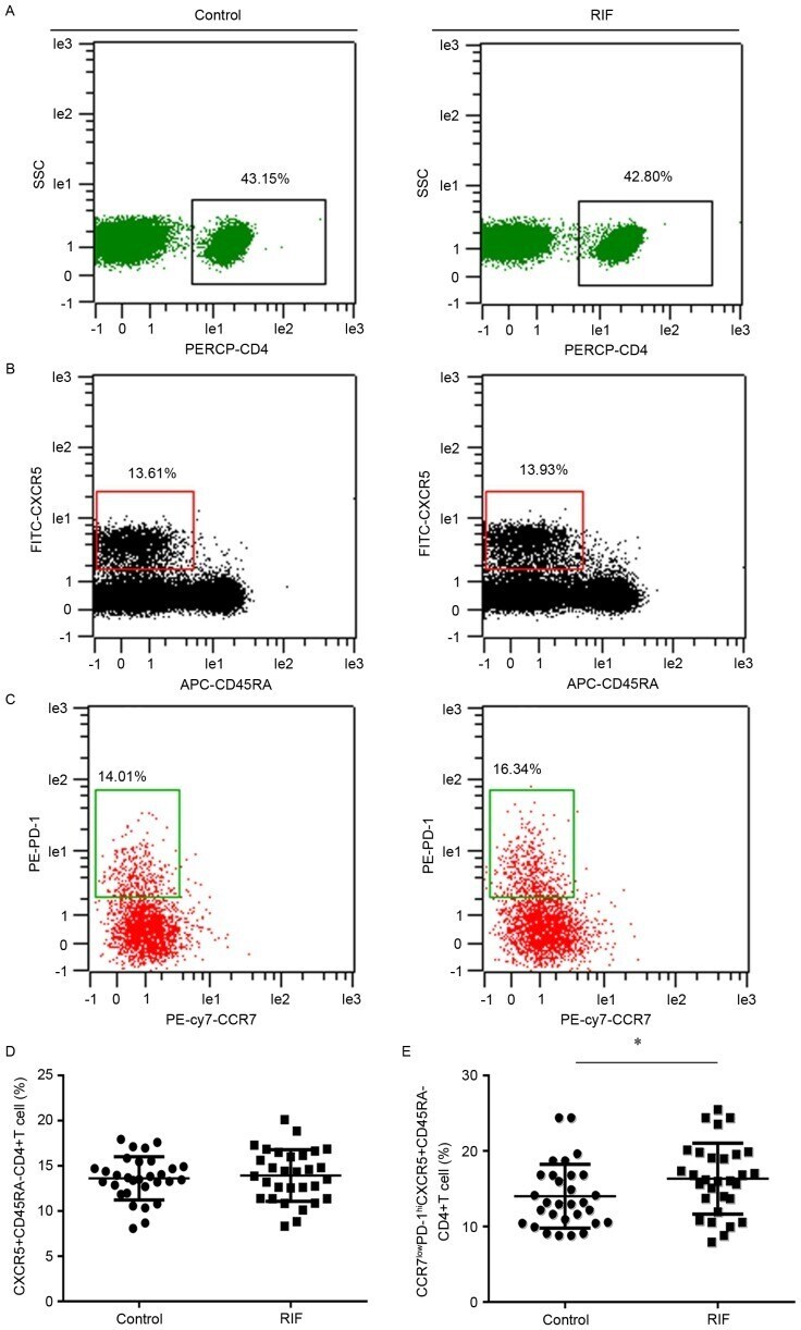
- Figure 1. Expression of CCR7(lo)PD-1(hi) T follicle helper cells in the peripheral blood. Cytometric plots indicating how (A) PERCP-CD4 was used to identify CD4 + T cells. These cells were further analyzed to identify cells expressing (B) CXCR5 and CD45RA. (C) CXCR5 + CD45RA - cells were analyzed to reveal the presence of CCR7 and PD-1. The presence of (D) CXCR5 + CD45RA - CD4 + T cells and (E) CCR7(lo)PD-1(hi) CXCR5 + CD45RA - CD4 + T cells in the RIF group and control group were compared. *P
- Conjugate
- Green dye
- Submitted by
- Invitrogen Antibodies (provider)
- Main image
- Experimental details
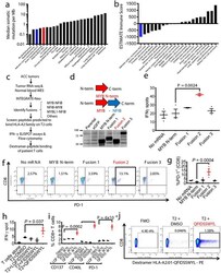
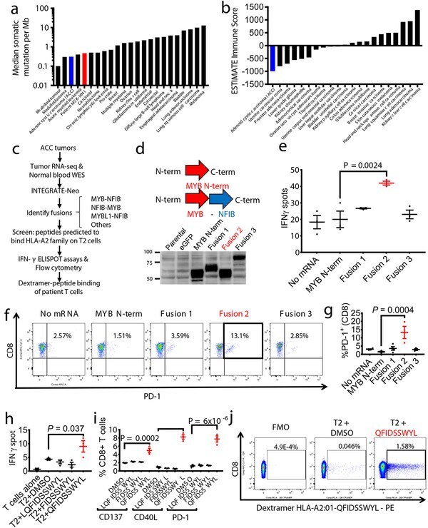
- Fig. 3 - MYB-NFIB generates an immuno-stimulatory peptide recognized by autologous T cells. a , Plot of median tumor mutational burden across 21 TCGA cancer types, adenoid cystic carcinomas 32 , 33 , and Patient MSK-HN1. b , Plot of mean ESTIMATE Immune Scores across 21 TCGA cancer types and adenoid cystic carcinomas. c , Experimental scheme for testing immunogenicity of MYB-NFIB fusion peptides from ACC patients. d , Diagram representation of the MYB-N-term and MYB-NFIB constructs expressed in HEK293 cells and validation by immunoblotting for MYB. Data are representative of two independent experiments. e , Quantification of IFN-gamma spots produced by patient T cells after 18 hr co-culture with healthy donor (""HD1""; HLA-A*02:01-positive) DCs (n=3) electroporated with mRNA. (one-way ANOVA followed by Dunnett's correction for multiple comparisons, 95%CI=-34.96 to -9.044, Effect size Eta Squared=0.782, DF=10, P =0.0024) f , Flow cytometry analysis of PD-1 expression on T cells used in (e). g , Quantification of CD3 + or CD8 + T cells used in (e) that are PD-1 + (n=3). (one-way ANOVA followed by Dunnett's correction for multiple comparisons, 95%CI=-17.58 to -5.877, Effect size Eta Squared=0.742, DF=13, P =0.0004) h , Quantification of IFN-gamma spots produced by patient T cells after 18 hr co-culture with T2 cells (n=3) pulsed with 10 muM of the indicated peptides. (one-way ANOVA followed by Dunnett's correction for multiple comparisons, 95%CI= -9.061 to -0.2724, Effect size E
- Conjugate
- Green dye
- Submitted by
- Invitrogen Antibodies (provider)
- Main image
- Experimental details
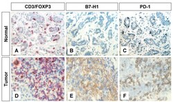
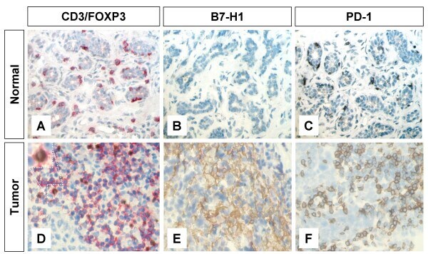
- Figure 1 Immunohistochemical staining of FOXP3+ T regs B7-H1 +and PD-1+ in T lymphocytes of breast tissues . Representative micrographs at x 530 magnification of (A&D) CD3/FOXP3 double staining (red color, membranous for CD3 and brown nuclear color for FOXP3 expression). (B&E) B7-H1 single staining (brown color, membranous/cytoplasmic expression). (C&F) PD-1 single staining (brown color, membranous expression). Upper panel (A-C) is sections for normal breast duct and lower panel (D-F) is sections for infiltrating ductal carcinoma of the breast.
- Conjugate
- Green dye
- Submitted by
- Invitrogen Antibodies (provider)
- Main image
- Experimental details
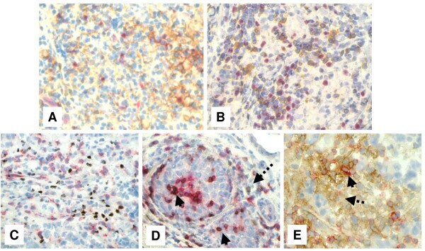
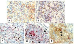
- Figure 4 Immunohistochemical staining showing the expression of FOXP3, B7-H1 and PD-1 molecules by different subsets of TIL . Representative micrographs at x 530 magnification of (A) double staining of B7-H1 (brown color, membranous/cytoplasmic) and FOXP3 (red color, nuclear) in an area rich in TIL of tumor section. (B) Double staining of PD-1 (brown color, membranous) and FOXP3 (red color, nuclear) in sections from the same tumor as in A. (C) double staining of FOXP3 (brown color, nuclear) and CD8 (red color, membranous). (D) double staining of PD-1 (brown color, membranous) and CD8 (red color, membranous). Solid arrows indicate CD8 + /PD-1 + T lymphocytes and dashed arrow indicates a CD8 - /PD-1 + T lymphocyte. (E) Double staining of B7-H1 (brown color, membranous/cytoplasmic) and CD8 (red color, membranous). Solid arrow indicates a CD8 + /B7-H1 + T lymphocyte and dashed arrow indicates a CD8 - /B7-H1 + T lymphocyte.
- Conjugate
- Green dye