 Explore
 Validate
 Learn
Learn Western blot
 Immunocytochemistry
 Immunohistochemistry
Antibody data
- Antibody Data
- Antigen structure
- References [3]
- Comments [0]
- Validations
- Immunocytochemistry [1]
Submit
Validation data
Reference
Comment
Report error
- Product number
- 14-2798-82 - Provider product page

- Provider
- Invitrogen Antibodies
- Product name
- CD279 (PD-1) Monoclonal Antibody (J121), eBioscience™
- Antibody type
- Monoclonal
- Antigen
- Other
- Description
- Description: This J121 monoclonal antibody reacts with human CD279 (programmed death-1, PD-1), a 55 kDa member of the CD28 immunoglobulin superfamily. CD279 contains the immunoreceptor tyrosine-based inhibitory motif (ITIM) and plays a key role in peripheral tolerance and autoimmune disease. CD279 is expressed predominantly on activated T and B lymphocytes. Two novel members of the B7 family have been identified as the CD279 ligands, CD274 (PD-L1, B7-H1) and CD273 (PD-L2, B7-DC). Evidence reported to date suggests overlapping functions for these two ligands and their constitutive expression on some normal tissues and upregulation on activated antigen-presenting cells. More recently, therapies targeting the blockade of the CD279/CD274 pathway have become the focus for treatment of melanoma, renal cell cancer, Hodgkins' lymphoma, and non-small cell lung carcinoma (NSCLC). The J121 monoclonal antibody is not recommended for flow cytometry of human cells. For detection of human CD279 using flow cytometry please refer to clone eBioJ105 (J105). Applications Reported: This J121 antibody has been reported for use in western blotting, and immunohistochemical staining of formalin-fixed paraffin embedded tissue sections. Applications Tested: This J121 antibody has been tested by immunohistochemistry of formalin-fixed paraffin embedded human tissue using high or low pH antigen retrieval and can be used at less than or equal to 5 µg/mL. It is recommended that the antibody be carefully titrated for optimal performance in the assay of interest. Purity: Greater than 90%, as determined by SDS-PAGE. Aggregation: Less than 10%, as determined by HPLC. Filtration: 0.2 µm post-manufacturing filtered.
- Reactivity
- Human
- Host
- Mouse
- Isotype
- IgG
- Antibody clone number
- J121
- Vial size
- 100 µg
- Concentration
- 0.5 mg/mL
- Storage
- 4° C
Submitted references T cell-NF-κB activation is required for tumor control in vivo.
PD-1 signaling in primary T cells.
Microanatomical localization of PD-1 in human tonsils.
Barnes SE, Wang Y, Chen L, Molinero LL, Gajewski TF, Evaristo C, Alegre ML
Journal for immunotherapy of cancer 2015;3(1):1
Journal for immunotherapy of cancer 2015;3(1):1
PD-1 signaling in primary T cells.
Riley JL
Immunological reviews 2009 May;229(1):114-25
Immunological reviews 2009 May;229(1):114-25
Microanatomical localization of PD-1 in human tonsils.
Iwai Y, Okazaki T, Nishimura H, Kawasaki A, Yagita H, Honjo T
Immunology letters 2002 Oct 1;83(3):215-20
Immunology letters 2002 Oct 1;83(3):215-20
No comments: Submit comment
Supportive validation
- Submitted by
- Invitrogen Antibodies (provider)
- Main image
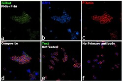
- Experimental details
- Immunofluorescence analysis of Programmed cell death protein 1 was performed using 70% confluent log phase Jurkat cells treated with PMA and PHA (50 ng/mL PHA and 1 mg/mL PMA, for 48 hours). The cells were fixed with 4% paraformaldehyde for 10 minutes, permeabilized with 0.1% Triton™ X-100 for 15 minutes, and blocked with 2% BSA for 45 minutes at room temperature. The cells were labeled with CD279 (PD-1) Monoclonal Antibody (J121), eBioscience™ (Product # 14-2798-82) at 1:100 dilution in 0.1% BSA, incubated at 4 degree celsius overnight and then labeled with Donkey anti-Mouse IgG (H+L) Highly Cross-Adsorbed Secondary Antibody, Alexa Fluor Plus 647 (Product # A32787), (1:2000 dilution), for 45 minutes at room temperature (Panel a: Green). Nuclei (Panel b:Blue) were stained with ProLong™ Diamond Antifade Mountant with DAPI (Product # P36962). F-actin (Panel c: Red) was stained with Rhodamine Phalloidin (Product # R415, 1:300). Panel d represents the merged image showing predominantly membrane localization. Panel e represents untreated Jurkat cells with low expression of CD279 (PD-1) Panel f represents control cells with no primary antibody to assess background. The images were captured at 60x magnification.
