 Explore
 Validate
 Learn
Learn Western blot
 Immunocytochemistry
Antibody data
- Antibody Data
- Antigen structure
- References [2]
- Comments [0]
- Validations
- Western blot [2]
- Immunohistochemistry [2]
Submit
Validation data
Reference
Comment
Report error
- Product number
- NBP2-24503 - Provider product page

- Provider
- Novus Biologicals
- Product name
- Rabbit Polyclonal ROR gamma/RORC/NR1F3 Antibody
- Antibody type
- Polyclonal
- Description
- Immunogen affinity purified. 100% homologous in human isoforms CRA_a).
- Reactivity
- Human
- Host
- Rabbit
- Isotype
- IgG
- Vial size
- 0.1 mg
- Concentration
- 1.0 mg/ml
- Storage
- Store at 4C short term. Aliquot and store at -20C long term. Avoid freeze-thaw cycles.
Submitted references Notch and the pre-TCR coordinate thymocyte proliferation by induction of the SCF subunits Fbxl1 and Fbxl12.
In vivo induction of cutaneous inflammation results in the accumulation of extracellular trap-forming neutrophils expressing RORγt and IL-17.
Zhao B, Yoganathan K, Li L, Lee JY, Zúñiga-Pflücker JC, Love PE
Nature immunology 2019 Oct;20(10):1381-1392
Nature immunology 2019 Oct;20(10):1381-1392
In vivo induction of cutaneous inflammation results in the accumulation of extracellular trap-forming neutrophils expressing RORγt and IL-17.
Keijsers RRMC, Hendriks AGM, van Erp PEJ, van Cranenbroek B, van de Kerkhof PCM, Koenen HJPM, Joosten I
The Journal of investigative dermatology 2014 May;134(5):1276-1284
The Journal of investigative dermatology 2014 May;134(5):1276-1284
No comments: Submit comment
Supportive validation
- Submitted by
- Novus Biologicals (provider)
- Main image
- Experimental details
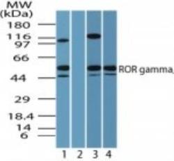
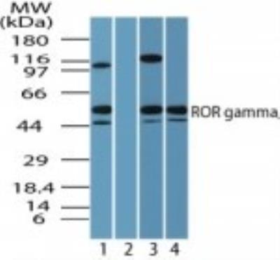
- Western Blot: ROR gamma/RORC/NR1F3 Antibody [NBP2-24503] - Analysis of human ROR gamma/RORC/NR1F3 in Jurkat cell lysate in the 1) absence, 2) presence of immunizing peptide, 3) in 3T3, and 4) in RAW cell lysate using NBP2-24503.
- Submitted by
- Novus Biologicals (provider)
- Main image

- Experimental details
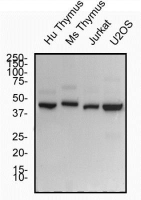
- Western Blot: ROR gamma/RORC/NR1F3 Antibody [NBP2-24503] - Total protein from Human and mouse Thymus, Jurkat and U2OS cells was separated on a 12% gel by SDS-PAGE, transferred to PVDF membrane and blocked in 5% non-fat milk in TBST. The membrane was probed with 2.0 ug/ml anti-ROR Gamma in 5% non-fat milk in TBST and detected with an anti-rabbit HRP secondary antibody using chemiluminescence.
Supportive validation
- Submitted by
- Novus Biologicals (provider)
- Main image
- Experimental details
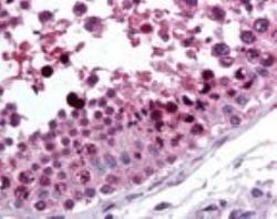
- Immunohistochemistry-Paraffin: ROR gamma/RORC/NR1F3 Antibody [NBP2-24503] - Analysis of human testis using NBP2-24503.

- Submitted by
- Novus Biologicals (provider)
- Main image
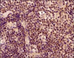
- Experimental details
- Immunohistochemistry-Paraffin: ROR gamma/RORC/NR1F3 Antibody [NBP2-24503] - Tissue section of human lymph node using anti- ROR gamma/RORC/NR1F3 antibody. The staining was developed with HRP labeled secondary antibody and DAB reagent, and nuclei of cells were counter-stained with hematoxylin. This antibody generated mainly a nuclear staining in a subset of cells and a weak to cytoplasmic staining was also observed in some cells.
