Explore
 Validate
 Learn
Learn Western blot
 Immunocytochemistry
Antibody data
- Antibody Data
- Antigen structure
- References [3]
- Comments [0]
- Validations
- Immunocytochemistry [1]
- Immunohistochemistry [1]
- Other assay [1]
Submit
Validation data
Reference
Comment
Report error
- Product number
- PA5-30583 - Provider product page

- Provider
- Invitrogen Antibodies
- Product name
- LOR Polyclonal Antibody
- Antibody type
- Polyclonal
- Antigen
- Synthetic peptide
- Description
- Recommended positive controls: Raji, HeLa. Store product as a concentrated solution. Centrifuge briefly prior to opening the vial.
- Reactivity
- Human, Mouse
- Host
- Rabbit
- Isotype
- IgG
- Vial size
- 100 μL
- Concentration
- 0.24 mg/mL
- Storage
- Store at 4°C short term. For long term storage, store at -20°C, avoiding freeze/thaw cycles.
Submitted references Lack of IRF6 Disrupts Human Epithelial Homeostasis by Altering Colony Morphology, Migration Pattern, and Differentiation Potential of Keratinocytes.
Unsaturated fatty acid-enriched extract from Hippophae rhamnoides seed reduces skin dryness through up-regulating aquaporins 3 and hyaluronan synthetases 2 expressions.
A Novel Van der Woude Syndrome-Causing IRF6 Variant Is Subject to Incomplete Non-sense-Mediated mRNA Decay Affecting the Phenotype of Keratinocytes.
Girousi E, Muerner L, Parisi L, Rihs S, von Gunten S, Katsaros C, Degen M
Frontiers in cell and developmental biology 2021;9:718066
Frontiers in cell and developmental biology 2021;9:718066
Unsaturated fatty acid-enriched extract from Hippophae rhamnoides seed reduces skin dryness through up-regulating aquaporins 3 and hyaluronan synthetases 2 expressions.
Yao Q, Jia T, Qiao W, Gu H, Kaku K
Journal of cosmetic dermatology 2021 Jan;20(1):321-329
Journal of cosmetic dermatology 2021 Jan;20(1):321-329
A Novel Van der Woude Syndrome-Causing IRF6 Variant Is Subject to Incomplete Non-sense-Mediated mRNA Decay Affecting the Phenotype of Keratinocytes.
Degen M, Girousi E, Feldmann J, Parisi L, La Scala GC, Schnyder I, Schaller A, Katsaros C
Frontiers in cell and developmental biology 2020;8:583115
Frontiers in cell and developmental biology 2020;8:583115
No comments: Submit comment
Supportive validation
- Submitted by
- Invitrogen Antibodies (provider)
- Main image
- Experimental details
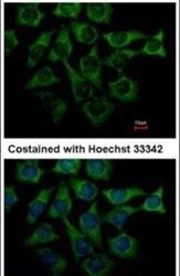
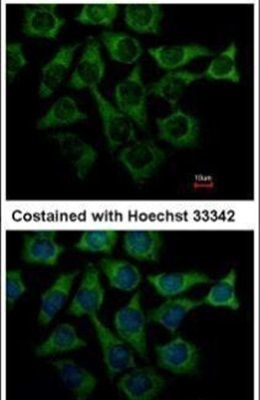
- Immunofluorescent analysis of Loricrin in methanol-fixed Hep3B cells using a Loricrin polyclonal antibody (Product # PA5-30583) at a 1:500 dilution.
Supportive validation
- Submitted by
- Invitrogen Antibodies (provider)
- Main image
- Experimental details
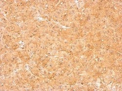
- LOR Polyclonal Antibody detects LOR protein at cytosol on HBL438 xenograft by immunohistochemical analysis. Sample: Paraffin-embedded HBL438 xenograft. LOR Polyclonal Antibody (Product # PA5-30583) dilution: 1:500. Antigen Retrieval: EDTA based buffer, pH 8.0, 15 min.

Supportive validation
- Submitted by
- Invitrogen Antibodies (provider)
- Main image
- Experimental details
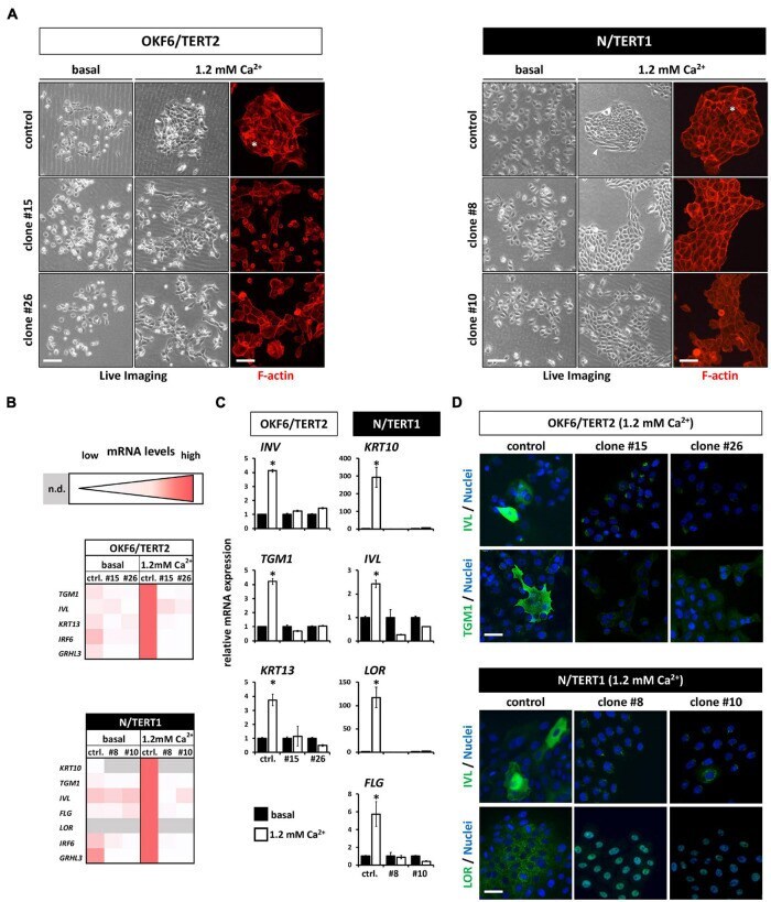
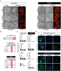
- FIGURE 5 (A) Live Imaging pictures and F-actin staining (phalloidin, red) with or without (only Live Imaging) exogenous Ca 2+ addition. While the control keratinocytes start to differentiate (asterisk and arrowheads), lack of IRF6 impairs differentiation. Scale bar: 200 mum (Live Imaging); Scale bar: 150 mum (F-actin). (B) Heatmaps of the qPCR analyses of various differentiation markers in basal (0.1 mM) vs. high Ca 2+ (1.2 mM) conditions. Note that in the absence of IRF6 all the differentiation markers are not induced upon the Ca 2+ -switch. n.d. : not detectable (Ct > 32); ctrl. : control. (C) qPCR analyses of specific differentiation markers showing the lack of induction upon the addition of Ca 2+ in the IRF6 knockout clones compared to controls. * p < 0.05 basal vs. 1.2 mM Ca 2+ . ctrl. : control. (D) Immunofluorescent staining for the markers Involucrin (IVL, green) and Transglutaminase 1 (TGM1, green) in OKF6/TERT2 cells and for IVL (green) and Loricrin (LOR, green) in N/TERT1 keratinocytes. Note that all differentiation markers were robustly induced in the control cells in the presence of IRF6. Scale bar: 50 mum. DAPI was used to counterstain the cell nuclei (blue).