 Explore
 Validate
 Learn
Learn Western blot
 Immunohistochemistry
Antibody data
- Antibody Data
- Antigen structure
- References [7]
- Comments [0]
- Validations
- Immunohistochemistry [1]
Submit
Validation data
Reference
Comment
Report error
- Product number
- HPA010553 - Provider product page

- Provider
- Atlas Antibodies
- Proper citation
- Atlas Antibodies Cat#HPA010553, RRID:AB_1853843
- Product name
- Anti-MFAP5
- Antibody type
- Polyclonal
- Description
- Polyclonal Antibody against Human MFAP5, Gene description: microfibrillar associated protein 5, Alternative Gene Names: MAGP2, MP25, Validated applications: IHC, WB, Uniprot ID: Q13361, Storage: Store at +4°C for short term storage. Long time storage is recommended at -20°C.
- Reactivity
- Human
- Host
- Rabbit
- Conjugate
- Unconjugated
- Isotype
- IgG
- Vial size
- 100 µl
- Concentration
- 0.2 mg/ml
- Storage
- Store at +4°C for short term storage. Long time storage is recommended at -20°C.
- Handling
- The antibody solution should be gently mixed before use.
Submitted references Highly efficient generation of self-renewing trophoblast from human pluripotent stem cells.
Microfibril Associated Protein 5 (MFAP5) Is Related to Survival of Ovarian Cancer Patients but Not Useful as a Prognostic Biomarker
Hypoxia-induced MFAP5 Promotes Tumor Migration and Invasion via AKT Pathway in Head and Neck Squamous Cell Carcinoma
Anticancer Immunotherapy by MFAP5 Blockade Inhibits Fibrosis and Enhances Chemosensitivity in Ovarian and Pancreatic Cancer
Spatial and Single-Cell Transcriptional Profiling Identifies Functionally Distinct Human Dermal Fibroblast Subpopulations.
MFAP5 and TNNC1: Potential markers for predicting occult cervical lymphatic metastasis and prognosis in early stage tongue cancer
Calcium-dependent FAK/CREB/TNNC1 signalling mediates the effect of stromal MFAP5 on ovarian cancer metastatic potential
Slamecka J, Ryu S, Tristan CA, Chu PH, Weber C, Deng T, Gedik Y, Ormanoglu P, Voss TC, Simeonov A, Singeç I
iScience 2024 Oct 18;27(10):110874
iScience 2024 Oct 18;27(10):110874
Microfibril Associated Protein 5 (MFAP5) Is Related to Survival of Ovarian Cancer Patients but Not Useful as a Prognostic Biomarker
Kujawa K, Zembala-Nożynska E, Syrkis J, Cortez A, Kupryjańczyk J, Lisowska K
International Journal of Molecular Sciences 2022;23(24):15994
International Journal of Molecular Sciences 2022;23(24):15994
Hypoxia-induced MFAP5 Promotes Tumor Migration and Invasion via AKT Pathway in Head and Neck Squamous Cell Carcinoma
Xu Q, Chang H, Tian X, Lou C, Ma H, Yang X
Journal of Cancer 2020;11(6):1596-1605
Journal of Cancer 2020;11(6):1596-1605
Anticancer Immunotherapy by MFAP5 Blockade Inhibits Fibrosis and Enhances Chemosensitivity in Ovarian and Pancreatic Cancer
Yeung T, Leung C, Yip K, Sheng J, Vien L, Bover L, Birrer M, Wong S, Mok S
Clinical Cancer Research 2019;25(21):6417-6428
Clinical Cancer Research 2019;25(21):6417-6428
Spatial and Single-Cell Transcriptional Profiling Identifies Functionally Distinct Human Dermal Fibroblast Subpopulations.
Philippeos C, Telerman SB, Oulès B, Pisco AO, Shaw TJ, Elgueta R, Lombardi G, Driskell RR, Soldin M, Lynch MD, Watt FM
The Journal of investigative dermatology 2018 Apr;138(4):811-825
The Journal of investigative dermatology 2018 Apr;138(4):811-825
MFAP5 and TNNC1: Potential markers for predicting occult cervical lymphatic metastasis and prognosis in early stage tongue cancer
Yang X, Wu K, Li S, Hu L, Han J, Zhu D, Tian X, Liu W, Tian Z, Zhong L, Yan M, Zhang C, Zhang Z
Oncotarget 2016;8(2):2525-2535
Oncotarget 2016;8(2):2525-2535
Calcium-dependent FAK/CREB/TNNC1 signalling mediates the effect of stromal MFAP5 on ovarian cancer metastatic potential
Leung C, Yeung T, Yip K, Pradeep S, Balasubramanian L, Liu J, Wong K, Mangala L, Armaiz-Pena G, Lopez-Berestein G, Sood A, Birrer M, Mok S
Nature Communications 2014;5(1)
Nature Communications 2014;5(1)
No comments: Submit comment
Supportive validation
- Submitted by
- Atlas Antibodies (provider)
- Enhanced method
- Orthogonal validation
- Main image
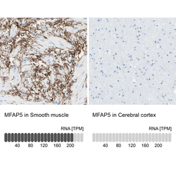
- Experimental details
- Immunohistochemistry analysis in human smooth muscle and cerebral cortex tissues using HPA010553 antibody. Corresponding MFAP5 RNA-seq data are presented for the same tissues.
- Sample type
- Human
- Protocol
- Protocol
