 Explore
 Validate
 Learn
LearnPA5-31553
antibody from Invitrogen Antibodies
Targeting: PYGO2
 Western blot
 Immunocytochemistry Immunoprecipitation Immunohistochemistry
 Chromatin Immunoprecipitation Other assay
Antibody data
- Antibody Data
- Antigen structure
- References [0]
- Comments [0]
- Validations
- Immunocytochemistry [2]
- Immunoprecipitation [1]
- Immunohistochemistry [2]
- Other assay [1]
Submit
Validation data
Reference
Comment
Report error
- Product number
- PA5-31553 - Provider product page

- Provider
- Invitrogen Antibodies
- Product name
- PYGO2 Polyclonal Antibody
- Antibody type
- Polyclonal
- Antigen
- Recombinant full-length protein
- Description
- Recommended positive controls: NIH-3T3. Predicted reactivity: Human (96%), Mouse (100%), Rat (99%), Pig (97%), Bovine (97%). Store product as a concentrated solution. Centrifuge briefly prior to opening the vial.
- Reactivity
- Human, Mouse, Rat
- Host
- Rabbit
- Isotype
- IgG
- Vial size
- 100 μL
- Concentration
- 0.65 mg/mL
- Storage
- Store at 4°C short term. For long term storage, store at -20°C, avoiding freeze/thaw cycles.
No comments: Submit comment
Supportive validation
- Submitted by
- Invitrogen Antibodies (provider)
- Main image
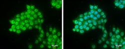
- Experimental details
- Immunocytochemistry-Immunofluorescence analysis of PYGO2 was performed in HCT 116 cells fixed in 4% paraformaldehyde at RT for 15 min. Green: PYGO2 Polyclonal Antibody (Product # PA5-31553) diluted at 1:500. Blue: Hoechst 33342 staining. Scale bar = 10 µm.

Supportive validation
- Submitted by
- Invitrogen Antibodies (provider)
- Main image
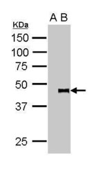
- Experimental details
- (Product # PA5-31553) IP Image. pygopus 2 was immunoprecipitated from 5*107 cells of SW480 whole cell lysate using 1 µg of anti-pygopus 2 antibody (Product # PA5-31553). The precipitated pygopus 2 was detected by (Product # PA5-31553) diluted at 1:15000. A: IP-IgG . Lane B: IP-pygopus 2 antibody.

Supportive validation
- Submitted by
- Invitrogen Antibodies (provider)
- Main image
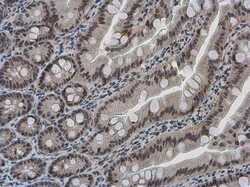
- Experimental details
- Immunohistochemistry (Paraffin) analysis of PYGO2 was performed in paraffin-embedded rat intestine tissue using PYGO2 Polyclonal Antibody (Product # PA5-31553) at a dilution of 1:500.
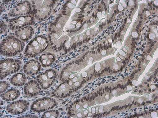
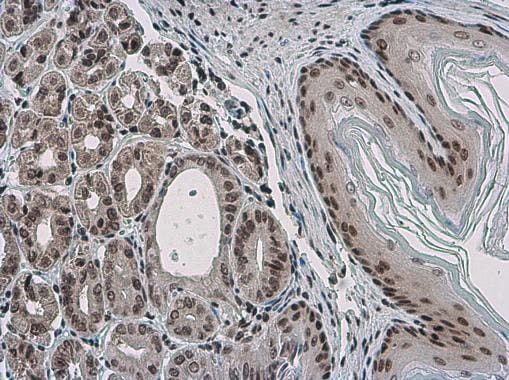
- Submitted by
- Invitrogen Antibodies (provider)
- Main image

- Experimental details
- Immunohistochemistry (Paraffin) analysis of PYGO2 was performed in paraffin-embedded mouse stomach tissue using PYGO2 Polyclonal Antibody (Product # PA5-31553) at a dilution of 1:500.
Supportive validation
- Submitted by
- Invitrogen Antibodies (provider)
- Main image

- Experimental details
- (Product # PA5-31553) IP Image. pygopus 2 was immunoprecipitated from 5*107 cells of SW480 whole cell lysate using 1 µg of anti-pygopus 2 antibody (Product # PA5-31553). The precipitated pygopus 2 was detected by (Product # PA5-31553) diluted at 1:15000. A: IP-IgG . Lane B: IP-pygopus 2 antibody.
