Explore
 Validate
 Learn
Learn Western blot
 Immunocytochemistry
Antibody data
- Antibody Data
- Antigen structure
- References [1]
- Comments [0]
- Validations
- Immunocytochemistry [2]
- Immunohistochemistry [1]
Submit
Validation data
Reference
Comment
Report error
- Product number
- MA5-24240 - Provider product page

- Provider
- Invitrogen Antibodies
- Product name
- PYGO2 Monoclonal Antibody (539730)
- Antibody type
- Monoclonal
- Antigen
- Recombinant full-length protein
- Description
- In direct ELISAs, no cross-reactivity with recombinant human Pygopus-1 or recombinant mouse Pygopus-1 is observed. Reconstitute in sterile PBS to a final concentration of 0.5 mg/mL.
- Reactivity
- Human
- Host
- Mouse
- Isotype
- IgG
- Antibody clone number
- 539730
- Vial size
- 100 μg
- Concentration
- 0.5 mg/mL
- Storage
- -20°C, Avoid Freeze/Thaw Cycles
Submitted references Discovery of an Orally Bioavailable Small-Molecule Inhibitor for the β-Catenin/B-Cell Lymphoma 9 Protein-Protein Interaction.
Wang Z, Zhang M, Quereda V, Frydman SM, Ming Q, Luca VC, Duckett DR, Ji H
Journal of medicinal chemistry 2021 Aug 26;64(16):12109-12131
Journal of medicinal chemistry 2021 Aug 26;64(16):12109-12131
No comments: Submit comment
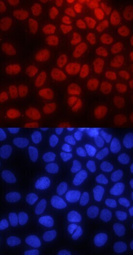
Supportive validation
- Submitted by
- Invitrogen Antibodies (provider)
- Main image

- Experimental details
- Immunocytochemistry analysis of PYGO2 in immersion fixed MCF‚7 human breast cancer cell line. Samples were incubated in PYGO2 monoclonal antibody (Product # MA5-24240) using a dilution of 10 µg/mL for 3 hours at room temperature followed by NorthernLights™ 557-conjugated Anti-Mouse IgG Secondary Antibody (red, upper panel) and counterstained with DAPI (blue, lower panel). Specific staining was localized to nuclei.

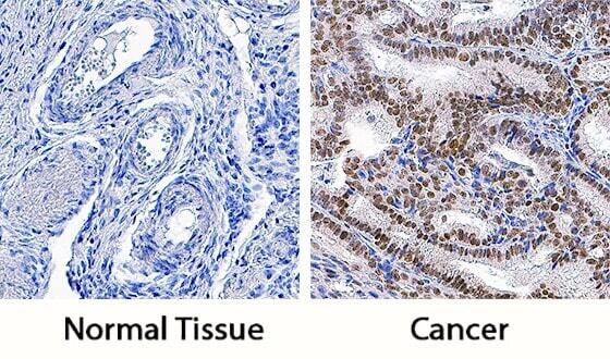
Supportive validation
- Submitted by
- Invitrogen Antibodies (provider)
- Main image
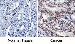
- Experimental details
- Immunohistochemical analysis of PYGO2 in immersion fixed paraffin-embedded sections of normal human ovary (left panel) and ovarian cancer tissue (right panel). Samples were incubated in PYGO2 monoclonal antibody (Product # MA5-24240) using a dilution of 3 µg/mL overnight at 4 °C. Tissue was stained using the Anti-Mouse HRP-DAB Cell & Tissue Staining Kit (brown) and counterstained with hematoxylin (blue). Specific staining was localized to nuclei.