Explore
 Validate
 Learn
LearnGTX119726
antibody from GeneTex
Targeting: PYGO2
 Western blot Immunocytochemistry Immunoprecipitation Immunohistochemistry
 Chromatin Immunoprecipitation
Antibody data
- Antibody Data
- Antigen structure
- References [3]
- Comments [0]
- Validations
- Western blot [2]
- Immunocytochemistry [1]
- Immunoprecipitation [1]
- Immunohistochemistry [3]
Submit
Validation data
Reference
Comment
Report error
- Product number
- GTX119726 - Provider product page

- Provider
- GeneTex
- Proper citation
- GeneTex Cat#GTX119726, RRID:AB_10618352
- Product name
- Pygopus 2 antibody
- Antibody type
- Polyclonal
- Reactivity
- Human, Mouse, Rat
- Host
- Rabbit
Submitted references An In Vivo Screen Identifies PYGO2 as a Driver for Metastatic Prostate Cancer.
lncRNA-dependent mechanisms of androgen-receptor-regulated gene activation programs.
Pygo2 regulates histone gene expression and H3 K56 acetylation in human mammary epithelial cells.
Lu X, Pan X, Wu CJ, Zhao D, Feng S, Zang Y, Lee R, Khadka S, Amin SB, Jin EJ, Shang X, Deng P, Luo Y, Morgenlander WR, Weinrich J, Lu X, Jiang S, Chang Q, Navone NM, Troncoso P, DePinho RA, Wang YA
Cancer research 2018 Jul 15;78(14):3823-3833
Cancer research 2018 Jul 15;78(14):3823-3833
lncRNA-dependent mechanisms of androgen-receptor-regulated gene activation programs.
Yang L, Lin C, Jin C, Yang JC, Tanasa B, Li W, Merkurjev D, Ohgi KA, Meng D, Zhang J, Evans CP, Rosenfeld MG
Nature 2013 Aug 29;500(7464):598-602
Nature 2013 Aug 29;500(7464):598-602
Pygo2 regulates histone gene expression and H3 K56 acetylation in human mammary epithelial cells.
Gu B, Watanabe K, Dai X
Cell cycle (Georgetown, Tex.) 2012 Jan 1;11(1):79-87
Cell cycle (Georgetown, Tex.) 2012 Jan 1;11(1):79-87
No comments: Submit comment
Supportive validation
- Submitted by
- GeneTex (provider)
- Main image
- Experimental details
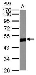
- Sample (30 ?g of whole cell lysate) A:NIH-3T310% SDS PAGE GTX119726 diluted at 1:1000 The HRP-conjugated anti-rabbit IgG antibody (GTX213110-01) was used to detect the primary antibody.

- Submitted by
- GeneTex (provider)
- Main image
- Experimental details
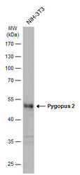
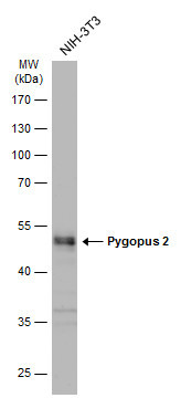
- Whole cell extract (30 ?g) was separated by 10% SDS-PAGE, and the membrane was blotted with Pygopus 2 antibody (GTX119726) diluted at 1:1000. The HRP-conjugated anti-rabbit IgG antibody (GTX213110-01) was used to detect the primary antibody.
Supportive validation
- Submitted by
- GeneTex (provider)
- Main image

- Experimental details
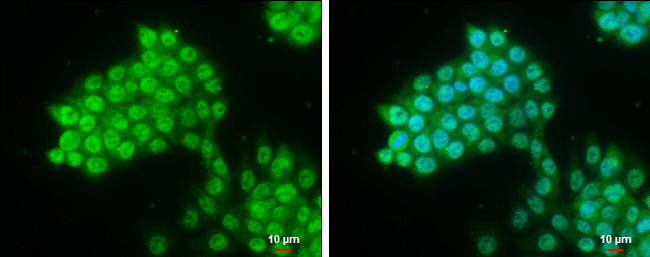
- Pygopus 2 antibody detects Pygopus 2 protein at cytoplasm and nucleus by immunofluorescent analysis.Sample: HCT 116 cells were fixed in 4% paraformaldehyde at RT for 15 min.Green: Pygopus 2 protein stained by Pygopus 2 antibody (GTX119726) diluted at 1:500.Blue: Hoechst 33342 staining.Scale bar = 10 £gm.
Supportive validation
- Submitted by
- GeneTex (provider)
- Main image

- Experimental details
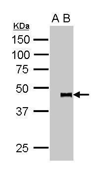
- GTX119726 IP Imagepygopus 2 was immunoprecipitated from 5*107 cells of SW480 whole cell lysate using 1 ug of anti-pygopus 2 antibody (GTX119726). The precipitated pygopus 2 was detected by GTX119726 diluted at 1:15000. A: IP-IgG B: IP-pygopus 2 antibody
Supportive validation
- Submitted by
- GeneTex (provider)
- Main image
- Experimental details
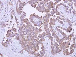
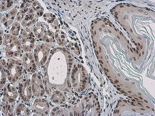
- pygopus 2 antibody detects Pygo2 protein at cytoplasm on human lung adenocarcinoma by immunohistochemical analysis. Sample: Paraffin-embedded lung adenocarcinoma. pygopus 2 antibody (GTX119726) dilution: 1:500.

- Submitted by
- GeneTex (provider)
- Main image
- Experimental details
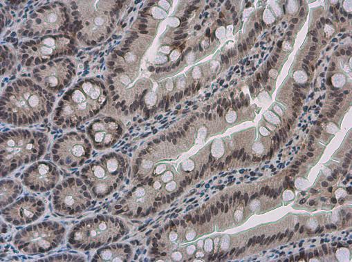
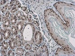
- Pygopus 2 antibody detects Pygopus 2 protein at nucleus in mouse stomach by immunohistochemical analysis. Sample: Paraffin-embedded mouse stomach. Pygopus 2 antibody (GTX119726) diluted at 1:500.
- Submitted by
- GeneTex (provider)
- Main image
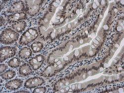
- Experimental details
- Pygopus 2 antibody detects Pygopus 2 protein at nucleus in rat intestine by immunohistochemical analysis. Sample: Paraffin-embedded rat intestine. Pygopus 2 antibody (GTX119726) diluted at 1:500.