Explore
 Validate
 Learn
Learn Western blot
 Immunocytochemistry
Antibody data
- Antibody Data
- Antigen structure
- References [2]
- Comments [0]
- Validations
- Immunocytochemistry [3]
- Immunohistochemistry [1]
- Other assay [4]
Submit
Validation data
Reference
Comment
Report error
- Product number
- PA5-21428 - Provider product page

- Provider
- Invitrogen Antibodies
- Product name
- SCAMP3 Polyclonal Antibody
- Antibody type
- Polyclonal
- Antigen
- Recombinant full-length protein
- Description
- Recommended positive controls: H1299, Molt-4. Predicted reactivity: Mouse (88%), Rat (83%), Pig (92%), Bovine (90%). Store product as a concentrated solution. Centrifuge briefly prior to opening the vial.
- Reactivity
- Human
- Host
- Rabbit
- Isotype
- IgG
- Vial size
- 100 μL
- Concentration
- 1 mg/mL
- Storage
- Store at 4°C short term. For long term storage, store at -20°C, avoiding freeze/thaw cycles.
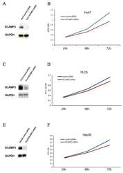
Submitted references SCAMP3 Regulates EGFR and Promotes Proliferation and Migration of Triple-Negative Breast Cancer Cells through the Modulation of AKT, ERK, and STAT3 Signaling Pathways.
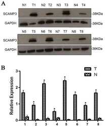
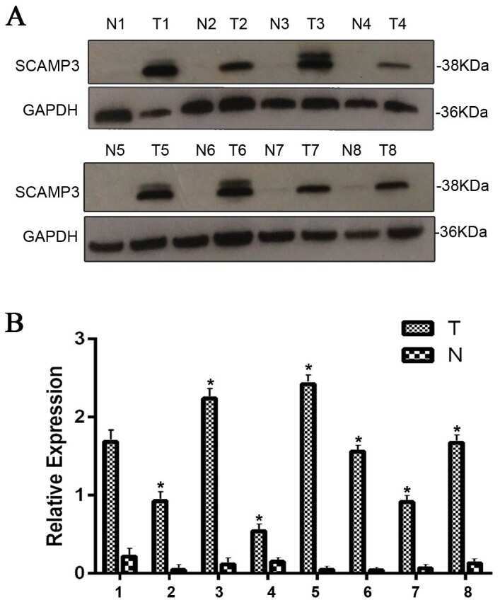
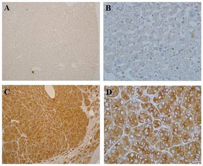
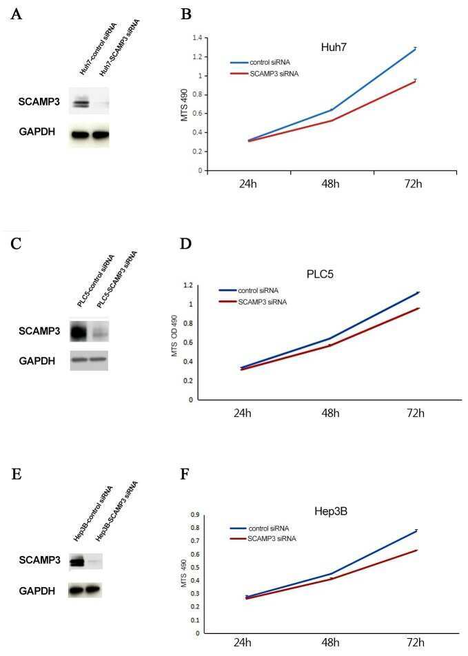
Overexpression of SCAMP3 is an indicator of poor prognosis in hepatocellular carcinoma.
Acevedo-Díaz A, Morales-Cabán BM, Zayas-Santiago A, Martínez-Montemayor MM, Suárez-Arroyo IJ
Cancers 2022 Jun 5;14(11)
Cancers 2022 Jun 5;14(11)
Overexpression of SCAMP3 is an indicator of poor prognosis in hepatocellular carcinoma.
Zhang X, Sheng J, Zhang Y, Tian Y, Zhu J, Luo N, Xiao C, Li R
Oncotarget 2017 Dec 12;8(65):109247-109257
Oncotarget 2017 Dec 12;8(65):109247-109257
No comments: Submit comment
Supportive validation
- Submitted by
- Invitrogen Antibodies (provider)
- Main image

- Experimental details
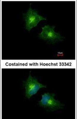
- Immunofluorescent analysis of SCAMP3 in methanol-fixed HeLa cells using a SCAMP3 polyclonal antibody (Product # PA5-21428) at a 1:1000 dilution.
- Submitted by
- Invitrogen Antibodies (provider)
- Main image
- Experimental details
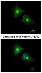
- Immunofluorescence analysis of methanol-fixed HeLa, using SCAMP3 antibody (Product # PA5-21428) at 1:1,000 dilution.

- Submitted by
- Invitrogen Antibodies (provider)
- Main image

- Experimental details
- Immunofluorescence analysis of methanol-fixed HeLa, using SCAMP3 antibody (Product # PA5-21428) at 1:1,000 dilution.

Supportive validation
- Submitted by
- Invitrogen Antibodies (provider)
- Main image
- Experimental details
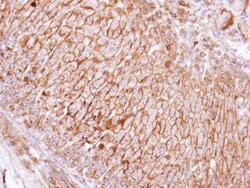
- Immunohistochemical analysis of paraffin-embedded human adrenal gland, using SCAMP3 (Product # PA5-21428) antibody at 1:100 dilution. Antigen Retrieval: EDTA based buffer, pH 8.0, 15 min.

Supportive validation
- Submitted by
- Invitrogen Antibodies (provider)
- Main image
- Experimental details
- NULL
- Submitted by
- Invitrogen Antibodies (provider)
- Main image

- Experimental details
- NULL
- Submitted by
- Invitrogen Antibodies (provider)
- Main image
- Experimental details
- NULL
- Submitted by
- Invitrogen Antibodies (provider)
- Main image

- Experimental details
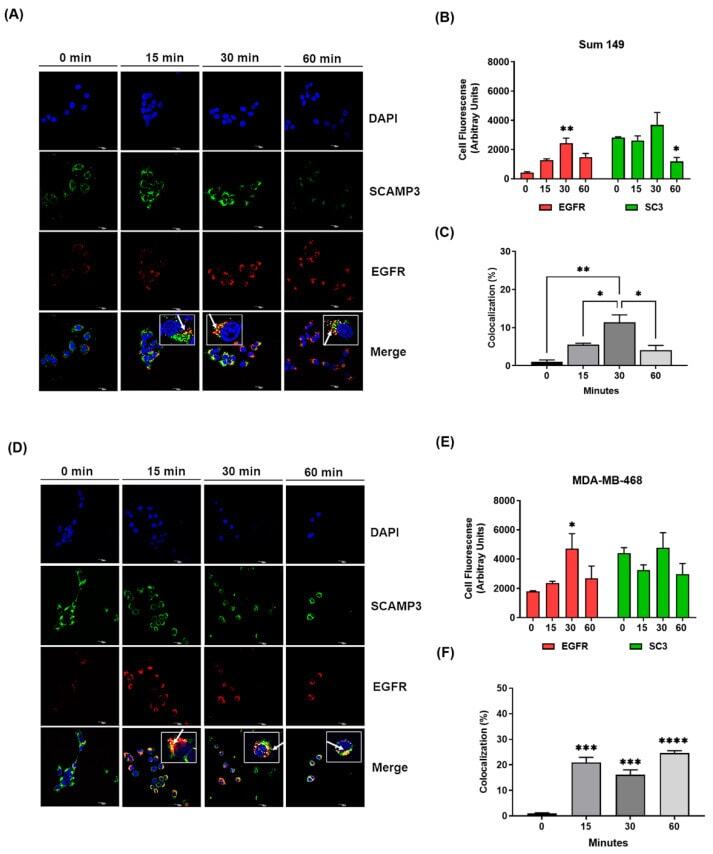
- Figure 4 SCAMP3 colocalizes with EGFR after receptor internalization. Cells were stimulated with 10 ng/mL EGF at the indicated time points to evaluate the location of SCAMP3 and EGFR using confocal microscopy. Representative images of the internalization assay and fluorescence quantification of SCAMP3, EGFR, and their colocalization in ( A - C ) SUM-149 and ( D - F ) MDA-MB-468 cells. The nuclei were stained with DAPI (blue). Secondary antibodies conjugated to Alexa 488 (green) and Alexa 594 (red) were used to detect SCAMP3 and EGFR, respectively. The micrographs were taken at a magnification of 60x using confocal microscopy. The white arrows indicate the colocalization of EGFR and SCAMP3 in the zoom images. The zoom-in of each image is shown in white squares and each has equal dimensions. Scale bar = 20 um. Total cell fluorescence and colocalization area analyses were performed in 20 cells from three experiments using Image J. Colocalization was calculated as the ratio of the colocalization fluorescence area to the total cell fluorescence area. One way or two-way ANOVA; * p