Explore
 Validate
 Learn
Learn Western blot
 Immunocytochemistry
Antibody data
- Antibody Data
- Antigen structure
- References [0]
- Comments [0]
- Validations
- Western blot [1]
- Immunohistochemistry [2]
Submit
Validation data
Reference
Comment
Report error
- Product number
- NBP2-12896 - Provider product page

- Provider
- Novus Biologicals
- Product name
- Mouse Monoclonal HCN3 Antibody
- Antibody type
- Monoclonal
- Description
- Protein G purified. Detects approx 90kDa. No cross-reactivity against other HCNs.
- Reactivity
- Human, Mouse, Rat
- Host
- Mouse
- Isotype
- IgG
- Vial size
- 0.1 mg
- Concentration
- 1 mg/ml
- Storage
- Store at 4C short term. Aliquot and store at -20C long term. Avoid freeze-thaw cycles.
No comments: Submit comment
Supportive validation
- Submitted by
- Novus Biologicals (provider)
- Main image
- Experimental details
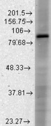
- Western Blot: HCN3 Antibody (S141-28) [NBP2-12896] - analysis of Rat brain membrane lysate showing detection of HCN3 protein using Mouse Anti-HCN3 Monoclonal Antibody, Clone S141-28 . Load: 15 ug protein. Block: 1.5% BSA for 30 minutes at RT. Primary Antibody: Mouse Anti-HCN3 Monoclonal Antibody at 1:1000 for 2 hours at RT. Secondary Antibody: Sheep Anti-Mouse IgG: HRP for 1 hour at RT.

Supportive validation
- Submitted by
- Novus Biologicals (provider)
- Main image
- Experimental details
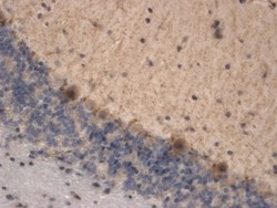
- Immunohistochemistry-Paraffin: HCN3 Antibody (S141-28) [NBP2-12896] - Tissue: brain tissue. Species: Mouse. Fixation: 10% Formalin Solution for 12-24 hours at RT. Primary Antibody: Mouse Anti-HCN3 Monoclonal Antibody at 1:1000 for 1 hour at RT. Secondary Antibody: HRP/DAB Detection System: Biotinylated Goat Anti-Mouse, Streptavidin Peroxidase, DAB Chromogen (brown) for 30 minutes at RT. Counterstain: Mayer Hematoxylin (purple/blue) nuclear stain at 250-500 ul for 5 minutes at RT. Localization: Granular staining of some Purkinje cells. Staining appears to be focal.

- Submitted by
- Novus Biologicals (provider)
- Main image
- Experimental details
- Immunohistochemistry-Paraffin: HCN3 Antibody (S141-28) [NBP2-12896] - Analysis of HCN3 in human hippocampal tissues using SMC-306. Courtesy of Dr. Turksen, Ottawa Hospital Research Institute, Canada.