Explore
 Validate
 Learn
LearnNBP2-21577
antibody from Novus Biologicals
Targeting: ARHGEF2
GEF-H1, GEFH1, KIAA0651, Lfc, LFP40, P40
 Western blot
 Immunocytochemistry
Antibody data
- Antibody Data
- Antigen structure
- References [0]
- Comments [0]
- Validations
- Western blot [10]
- Immunohistochemistry [6]
Submit
Validation data
Reference
Comment
Report error
- Product number
- NBP2-21577 - Provider product page

- Provider
- Novus Biologicals
- Product name
- Rabbit Polyclonal GEF-H1 Antibody
- Antibody type
- Polyclonal
- Description
- Immunogen affinity purified.
- Reactivity
- Human, Mouse, Rat
- Host
- Rabbit
- Isotype
- IgG
- Vial size
- 0.1 ml
- Storage
- Aliquot and store at -20C or -80C. Avoid freeze-thaw cycles.
No comments: Submit comment
Supportive validation
- Submitted by
- Novus Biologicals (provider)
- Main image
- Experimental details
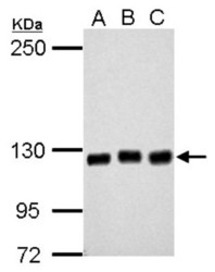
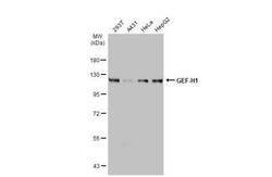
- Western Blot: GEF-H1 Antibody [NBP2-21577] - Sample (30 ug of whole cell lysate) A: 293T B: A431 C: HeLa 5% SDS PAGE gel, diluted at 1:2000.
- Submitted by
- Novus Biologicals (provider)
- Main image
- Experimental details
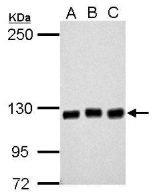
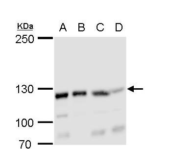
- Western Blot: GEF-H1 Antibody [NBP2-21577] - A. 30 ug Raw264. 7 whole cell lysate/extract B. 30 ug C2C12 whole cell lysate/extract 5 % SDS-PAGE GEF-H1 antibody dilution: 1:1000. (Mouse)
- Submitted by
- Novus Biologicals (provider)
- Main image
- Experimental details
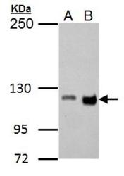
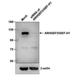
- Western Blot: GEF-H1 Antibody [NBP2-21577] - Sample (10 ug of whole cell lysate) A: hMSC-3A6 B: siRNA of ARHGEF2/GEF-H1 8% SDS PAGE diluted at 1:1000. The HRP-conjugated anti-rabbit IgG antibody (NBP2-19301) was used to detect the primary antibody.

- Submitted by
- Novus Biologicals (provider)
- Main image
- Experimental details
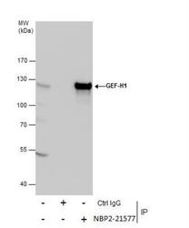
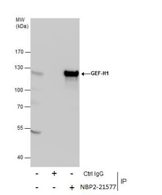
- Western Blot: GEF-H1 Antibody [NBP2-21577] - Immunoprecipitation of GEF-H1 protein from 293T whole cell extracts using 5 ug of GEF-H1 antibody. Western blot analysis was performed using GEF-H1 antibody. EasyBlot anti-Rabbit IgG was used as a secondary reagent.
- Submitted by
- Novus Biologicals (provider)
- Main image
- Experimental details
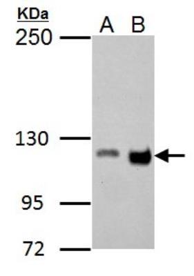
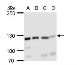
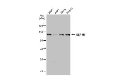
- Western Blot: GEF-H1 Antibody [NBP2-21577] - A. 30 ug 293T whole cell lysate/extract B. 30 ug A431 whole cell lysate/extract C. 30 ug HeLa whole cell lysate/extract D. 30 ug HepG2 whole cell lysate/extract 5% SDS-PAGE GEF-H1 antibody dilution: 1:2000 The HRP-conjugated anti-rabbit IgG antibody (NBP2-19301) was used to detect the primary antibody.

- Submitted by
- Novus Biologicals (provider)
- Main image
- Experimental details
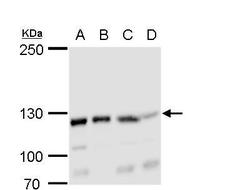
- Western Blot: GEF-H1 Antibody [NBP2-21577] - A. 30 ug 293T whole cell lysate/extract B. 30 ug A431 whole cell lysate/extract C. 30 ug HeLa whole cell lysate/extract D. 30 ug HepG2 whole cell lysate/extract 5% SDS-PAGE GEF-H1 antibody dilution: 1:2000. The HRP-conjugated anti-rabbit IgG antibody (NBP2-19301) was used to detect the primary antibody.
- Submitted by
- Novus Biologicals (provider)
- Main image
- Experimental details
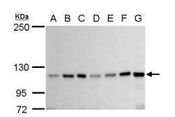
- Western Blot: GEF-H1 Antibody [NBP2-21577] - A. 30 ug Neuro2A whole cell lysate/extract B. 30 ug GL261 whole cell lysate/extract C. 30 ug C8D30 whole cell lysate/extract D. 30 ug NIH-3T3 whole cell lysate/extract E. 30 ug BCL-1 whole cell lysate/extract F. 30 ug Raw 264.7 whole cell lysate/extract G. 30 ug C2Cl2 whole cell lysate/extract 5% SDS-PAGE GEF-H1 antibody dilution: 1:1000. The HRP-conjugated anti-rabbit IgG antibody (NBP2-19301) was used to detect the primary antibody.

- Submitted by
- Novus Biologicals (provider)
- Main image
- Experimental details
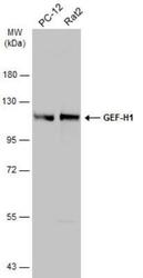
- Western Blot: GEF-H1 Antibody [NBP2-21577] - Various whole cell extracts (30 ug) were separated by 7.5% SDS-PAGE, and the membrane was blotted with GEF-H1 antibody diluted at 1:500. The HRP-conjugated anti-rabbit IgG antibody (NBP2-19301) was used to detect the primary antibody.
- Submitted by
- Novus Biologicals (provider)
- Main image
- Experimental details
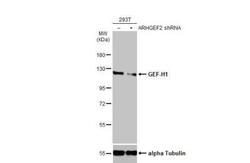
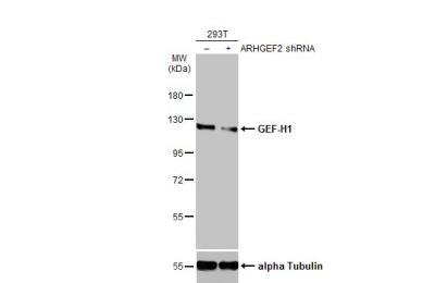
- Western Blot: GEF-H1 Antibody [NBP2-21577] - Non-transfected (-) and transfected (+) 293T whole cell extracts (30 ug) were separated by 7.5% SDS-PAGE, and the membrane was blotted with GEF-H1 antibody diluted at 1:4000. HRP-conjugated anti-rabbit IgG antibody was used to detect the primary antibody.
- Submitted by
- Novus Biologicals (provider)
- Main image
- Experimental details
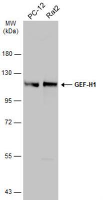
- Western Blot: GEF-H1 Antibody [NBP2-21577] - Various whole cell extracts (30 ug) were separated by 7.5% SDS-PAGE, and the membrane was blotted with GEF-H1 antibody diluted at 1:2000. HRP-conjugated anti-rabbit IgG antibody was used to detect the primary antibody.
Supportive validation
- Submitted by
- Novus Biologicals (provider)
- Main image

- Experimental details
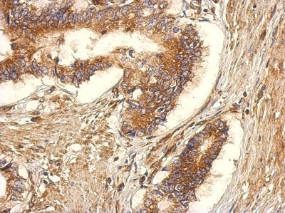
- Immunohistochemistry-Paraffin: GEF-H1 Antibody [NBP2-21577] - Immunohistochemical analysis of paraffin-embedded Gastric ca, using antibody at 1:500 dilution.
- Submitted by
- Novus Biologicals (provider)
- Main image
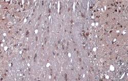
- Experimental details
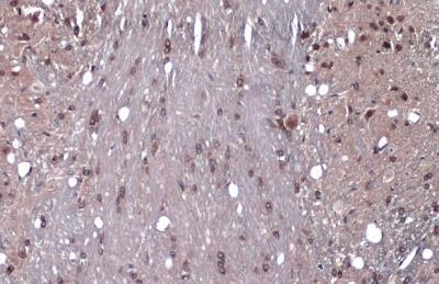
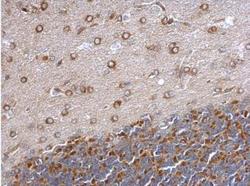
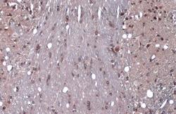
- Immunohistochemistry-Paraffin: GEF-H1 Antibody [NBP2-21577] - Paraffin-embedded rat hind brain. GEF-H1 antibody dilution: 1:500.

- Submitted by
- Novus Biologicals (provider)
- Main image
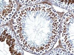
- Experimental details
- Immunohistochemistry-Paraffin: GEF-H1 Antibody [NBP2-21577] - Paraffin-embedded mouse testis. GEF-H1 antibody dilution: 1:500.

- Submitted by
- Novus Biologicals (provider)
- Main image
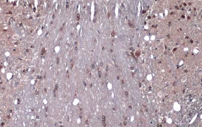
- Experimental details
- Immunohistochemistry-Paraffin: GEF-H1 Antibody [NBP2-21577] - mouse brain. GEF-H1 stained by GEF-H1 antibody diluted at 1:1000. Antigen Retrieval: Citrate buffer, pH 6.0, 15 min
- Submitted by
- Novus Biologicals (provider)
- Main image
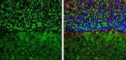
- Experimental details
- Immunohistochemistry-Frozen: GEF-H1 Antibody [NBP2-21577] - Frozen-sectioned mouse mouse cerebellum. Green: GEF-H1 stained by GEF-H1 antibody diluted at 1:250. Red: NF-H, stained by NF-H antibody [114] diluted at 1:500. Blue: Fluoroshield with DAPI. Antigen Retrieval: Citrate buffer, pH 6.0, 10 min.

- Submitted by
- Novus Biologicals (provider)
- Main image
- Experimental details
- Immunohistochemistry-Paraffin: GEF-H1 Antibody [NBP2-21577] - GEF-H1 antibody detects GEF-H1 protein at cytoplasm by immunohistochemical analysis. Sample: Paraffin-embedded mouse brain. GEF-H1 stained by GEF-H1 antibody diluted at 1:1000. Antigen Retrieval: Citrate buffer, pH 6.0, 15 min.