Explore
 Validate
 Learn
Learn Western blot
 Immunocytochemistry
Antibody data
- Antibody Data
- Antigen structure
- References [3]
- Comments [0]
- Validations
- Western blot [6]
- Immunocytochemistry [1]
- Immunoprecipitation [2]
- Immunohistochemistry [3]
Submit
Validation data
Reference
Comment
Report error
- Product number
- GTX125893 - Provider product page

- Provider
- GeneTex
- Proper citation
- GeneTex Cat#GTX125893, RRID:AB_11177391
- Product name
- GEF-H1 antibody
- Antibody type
- Polyclonal
- Reactivity
- Human, Mouse, Rat
- Host
- Rabbit
Submitted references The pseudokinase CaMKv is required for the activity-dependent maintenance of dendritic spines.
Dexamethasone-induced cellular tension requires a SGK1-stimulated Sec5-GEF-H1 interaction.
GEF-H1 controls focal adhesion signaling that regulates mesenchymal stem cell lineage commitment.
Liang Z, Zhan Y, Shen Y, Wong CC, Yates JR 3rd, Plattner F, Lai KO, Ip NY
Nature communications 2016 Oct 31;7:13282
Nature communications 2016 Oct 31;7:13282
Dexamethasone-induced cellular tension requires a SGK1-stimulated Sec5-GEF-H1 interaction.
Wang HL, Yang CH, Lee HH, Kuo JC, Hur SS, Chien S, Lee OK, Hung SC, Chang ZF
Journal of cell science 2015 Oct 15;128(20):3757-68
Journal of cell science 2015 Oct 15;128(20):3757-68
GEF-H1 controls focal adhesion signaling that regulates mesenchymal stem cell lineage commitment.
Huang IH, Hsiao CT, Wu JC, Shen RF, Liu CY, Wang YK, Chen YC, Huang CM, del Álamo JC, Chang ZF, Tang MJ, Khoo KH, Kuo JC
Journal of cell science 2014 Oct 1;127(Pt 19):4186-200
Journal of cell science 2014 Oct 1;127(Pt 19):4186-200
No comments: Submit comment
Enhanced validation
Supportive validation
- Submitted by
- GeneTex (provider)
- Enhanced method
- Genetic validation
- Main image
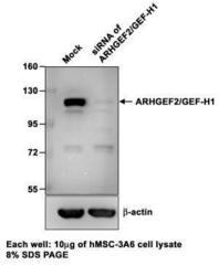
- Experimental details
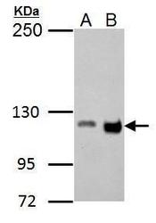
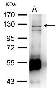
- Sample (10 ?g of whole cell lysate) A: hMSC-3A6 B: siRNA of ARHGEF2/GEF-H1 8% SDS PAGE GTX125893 diluted at 1:1000 The HRP-conjugated anti-rabbit IgG antibody (GTX213110-01) was used to detect the primary antibody.

Supportive validation
- Submitted by
- GeneTex (provider)
- Main image
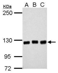
- Experimental details
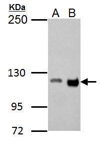
- Sample (30 ?g of whole cell lysate) A: 293T B: A431 C: HeLa 5% SDS PAGE GTX125893 diluted at 1:2000 The HRP-conjugated anti-rabbit IgG antibody (GTX213110-01) was used to detect the primary antibody.
- Submitted by
- GeneTex (provider)
- Main image
- Experimental details
- GEF-H1 antibody detects GEF-H1 protein by western blot analysis.A. 30 ?g Raw264.7 whole cell lysate/extractB. 30 ?g C2C12 whole cell lysate/extract5% SDS-PAGEGEF-H1 antibody (GTX125893) dilution: 1:1000 The HRP-conjugated anti-rabbit IgG antibody (GTX213110-01) was used to detect the primary antibody.
- Submitted by
- GeneTex (provider)
- Main image

- Experimental details
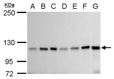
- GEF-H1 antibody detects GEF-H1 protein by western blot analysis.A. 30 ?g Neuro2A whole cell lysate/extractB. 30 ?g GL261 whole cell lysate/extract C. 30 ?g C8D30 whole cell lysate/extract D. 30 ?g NIH-3T3 whole cell lysate/extract E. 30 ?g BCL-1 whole cell lysate/extract F. 30 ?g Raw 264.7 whole cell lysate/extract G. 30 ?g C2Cl2 whole cell lysate/extract5% SDS-PAGEGEF-H1 antibody (GTX125893) dilution: 1:1000The HRP-conjugated anti-rabbit IgG antibody (GTX213110-01) was used to detect the primary antibody.
- Submitted by
- GeneTex (provider)
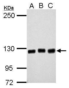
- Main image
- Experimental details
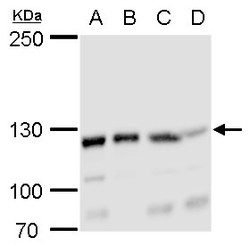
- GEF-H1 antibody detects GEF-H1 protein by western blot analysis.A. 30 ?g 293T whole cell lysate/extract B. 30 ?g A431 whole cell lysate/extract C. 30 ?g HeLa whole cell lysate/extract D. 30 ?g HepG2 whole cell lysate/extract5% SDS-PAGEGEF-H1 antibody (GTX125893) dilution: 1:2000The HRP-conjugated anti-rabbit IgG antibody (GTX213110-01) was used to detect the primary antibody.

- Submitted by
- GeneTex (provider)
- Main image

- Experimental details
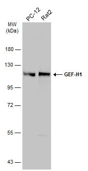
- Various whole cell extracts (30 ?g) were separated by 7.5% SDS-PAGE, and the membrane was blotted with GEF-H1 antibody (GTX125893) diluted at 1:500. The HRP-conjugated anti-rabbit IgG antibody (GTX213110-01) was used to detect the primary antibody.
Supportive validation
- Submitted by
- GeneTex (provider)
- Main image

- Experimental details
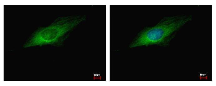
- GEF-H1 antibody detects ARHGEF2 protein at Cytoplasm by immunofluorescent analysis. Sample: HeLa cells were fixed in -20¢J 100% MeOH for 5 min.Green: ARHGEF2 protein stained by GEF-H1 antibody (GTX125893) diluted at 1:500.Blue: Hoechst 33343 staining.
Supportive validation
- Submitted by
- GeneTex (provider)
- Main image

- Experimental details
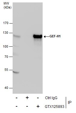
- GEF H1 antibody immunoprecipitates GEF H1 protein in IP experiments.IP Sample: 700 µg 3A6 whole cell lysate/extract A. Immunoprecipitation of EXOSC 10 protein by 3 ?g of GEF H1 antibody (GTX125893)8% SDS-PAGE
- Validation comment
- IP
- Submitted by
- GeneTex (provider)
- Main image
- Experimental details
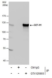
- Immunoprecipitation of GEF-H1 protein from 293T whole cell extracts using 5 £gg of GEF-H1 antibody (GTX125893).Western blot analysis was performed using GEF-H1 antibody (GTX125893).EasyBlot anti-Rabbit IgG (GTX221666-01) was used as a secondary reagent.
Supportive validation
- Submitted by
- GeneTex (provider)
- Main image
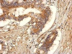
- Experimental details
- Immunohistochemical analysis of paraffin-embedded human gastric cancer, using GEF-H1(GTX125893) antibody at 1:500 dilution.
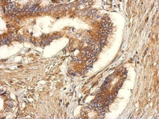
- Submitted by
- GeneTex (provider)
- Main image
- Experimental details
- GEF-H1 antibody detects GEF-H1 protein at cytosol on mouse testis by immunohistochemical analysis. Sample: Paraffin-embedded mouse testis. GEF-H1 antibody (GTX125893) dilution: 1:500.

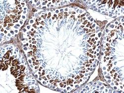
- Submitted by
- GeneTex (provider)
- Main image
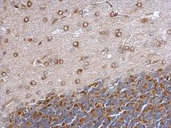
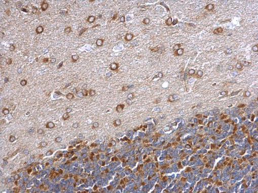
- Experimental details
- GEF-H1 antibody detects GEF-H1 protein at cytosol on rat hind brain by immunohistochemical analysis. Sample: Paraffin-embedded rat hind brain. GEF-H1 antibody (GTX125893) dilution: 1:500.