 Explore
 Validate
 Learn
LearnNBP2-76838
antibody from Novus Biologicals
Targeting: ARHGEF2
GEF-H1, GEFH1, KIAA0651, Lfc, LFP40, P40
 Western blot
 Immunocytochemistry
Antibody data
- Antibody Data
- Antigen structure
- References [0]
- Comments [0]
- Validations
- Western blot [1]
- Immunohistochemistry [3]
- Flow cytometry [1]
Submit
Validation data
Reference
Comment
Report error
- Product number
- NBP2-76838 - Provider product page

- Provider
- Novus Biologicals
- Product name
- Rabbit Monoclonal GEF-H1 Antibody
- Antibody type
- Monoclonal
- Description
- Protein A purified.
- Reactivity
- Human, Mouse, Rat
- Host
- Rabbit
- Isotype
- IgG
- Vial size
- 100 ul
- Storage
- Store at 4C short term. Aliquot and store at -20C long term. Avoid freeze-thaw cycles.
No comments: Submit comment
Supportive validation
- Submitted by
- Novus Biologicals (provider)
- Main image
- Experimental details
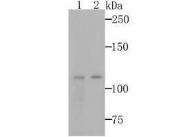
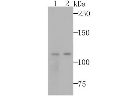
- Western Blot: GEF-H1 Antibody (JG36-46) [NBP2-76838] - Western blot analysis of GEF H1 on 293T and A431 cells lysates using anti-GEF H1 antibody at 1/500 dilution.
Supportive validation
- Submitted by
- Novus Biologicals (provider)
- Main image
- Experimental details
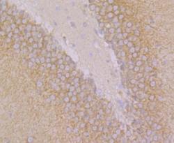
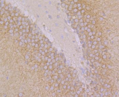
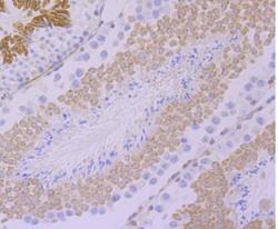
- Immunohistochemistry: GEF-H1 Antibody (JG36-46) [NBP2-76838] - Immunohistochemical analysis of paraffin-embedded rat brain tissue using anti-GEF H1 antibody. Counter stained with hematoxylin.
- Submitted by
- Novus Biologicals (provider)
- Main image
- Experimental details
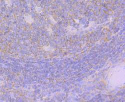
- Immunohistochemistry: GEF-H1 Antibody (JG36-46) [NBP2-76838] - Immunohistochemical analysis of paraffin-embedded human tonsil tissue using anti-GEF H1 antibody. Counter stained with hematoxylin.
- Submitted by
- Novus Biologicals (provider)
- Main image
- Experimental details
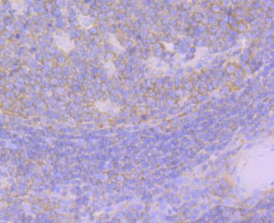
- Immunohistochemistry: GEF-H1 Antibody (JG36-46) [NBP2-76838] - Immunohistochemical analysis of paraffin-embedded mouse testis tissue using anti-GEF H1 antibody. Counter stained with hematoxylin.

Supportive validation
- Submitted by
- Novus Biologicals (provider)
- Main image
- Experimental details
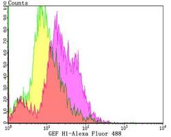
- Flow Cytometry: GEF-H1 Antibody (JG36-46) [NBP2-76838] - Flow cytometric analysis of HL-60 cells with GEF H1 antibody at 1/100 dilution (yellow) compared with an unlabelled control (cells without incubation with primary antibody; purple).Alexa Fluor 488-conjugated goat anti-rabbit IgG was used as the secondary antibody.
