 Explore
 Validate
 Learn
Learn Western blot
Antibody data
- Antibody Data
- Antigen structure
- References [0]
- Comments [0]
- Validations
- Western blot [1]
- Immunocytochemistry [1]
Submit
Validation data
Reference
Comment
Report error
- Product number
- MA3-017 - Provider product page

- Provider
- Invitrogen Antibodies
- Product name
- CRABP2 Monoclonal Antibody (5CRA-3B3)
- Antibody type
- Monoclonal
- Antigen
- Recombinant full-length protein
- Description
- MA3-017 has been used successfully in Western blotting and Immunofluorescence. This antibody was referenced in publications to perform in IHC.
- Reactivity
- Human
- Host
- Mouse
- Isotype
- IgG
- Antibody clone number
- 5CRA-3B3
- Vial size
- 50 µL
- Concentration
- Conc. Not Determined
- Storage
- -20° C, Avoid Freeze/Thaw Cycles
No comments: Submit comment
Supportive validation
- Submitted by
- Invitrogen Antibodies (provider)
- Main image

- Experimental details
- Western blot analysis of CRABP2 was performed by loading 50 µg of the indicated whole cell lysates and 5 µL of PageRuler Plus Prestained Protein Ladder (Product # 26619) per well onto a Novex 4-20% Tris-Glycine polyacrylamide gel (Product # WT4202BOX ). Proteins were transferred to a PVDF membrane using the G2 Blotter (Product # 62288), and blocked with Starting Block T20 (Product # 37543) for 1 hour at room temperature. CRABP2 was detected at ~14 kD using a CRABP2 monoclonal antibody (Product # MA3-017) at a dilution of 1:2000 in blocking buffer for 1 hour at room temperature on a rocking platform, followed by a Goat anti-Mouse IgG Secondary Antibody, HRP conjugate (Product # 31430) at a dilution of 1:20,000 for at least 30 minutes at room temperature. Chemiluminescent detection was performed using SuperSignal Pico substrate (Product # 34078) and the myECL Imager (Product # 62236).

Supportive validation
- Submitted by
- Invitrogen Antibodies (provider)
- Main image
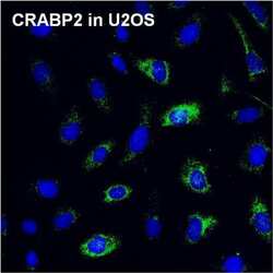
- Experimental details
- Immunofluorescent analysis of CRABP2 (green) in U2OS cells. The cells were fixed with 4% paraformaldehyde for 15 minutes, permeabilized with 0.1% Triton X-100 in PBS for 10 minutes, and blocked with 3% BSA in PBS (Product # 37525) for 30 minutes at room temperature. Cells were stained with a CRABP2 monoclonal antibody (Product # MA3-017) at a dilution of 1:100 in staining buffer for 1 hour at room temperature, and then incubated with a Goat anti-Mouse IgG Secondary Antibody, DyLight 488 conjugate (Product # 35502) at a dilution of 1:500 for 1 hour at room temperature (green). Nuclei (blue) were stained with Hoechst 33342 dye (Product # 62249). Images were taken on a Thermo Scientific ToxInsight Instrument at 20X magnification.
