 Explore
 Validate
 Learn
Learn Western blot
 Immunocytochemistry
Antibody data
- Antibody Data
- Antigen structure
- References [2]
- Comments [0]
- Validations
- Immunocytochemistry [2]
- Immunohistochemistry [2]
- Other assay [2]
Submit
Validation data
Reference
Comment
Report error
- Product number
- PA5-27451 - Provider product page

- Provider
- Invitrogen Antibodies
- Product name
- CRABP2 Polyclonal Antibody
- Antibody type
- Polyclonal
- Antigen
- Recombinant full-length protein
- Description
- Recommended positive controls: A549, HepG2, mouse brain, mouse fetal brain, rat brain. Predicted reactivity: Mouse (94%), Rat (94%), Xenopus laevis (89%), Pig (95%), Rhesus Monkey (100%), Bovine (97%). Store product as a concentrated solution. Centrifuge briefly prior to opening the vial.
- Reactivity
- Human, Mouse, Rat
- Host
- Rabbit
- Isotype
- IgG
- Vial size
- 100 μL
- Concentration
- 1 mg/mL
- Storage
- Store at 4°C short term. For long term storage, store at -20°C, avoiding freeze/thaw cycles.
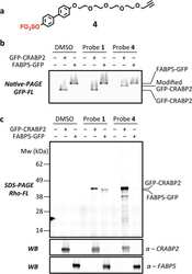
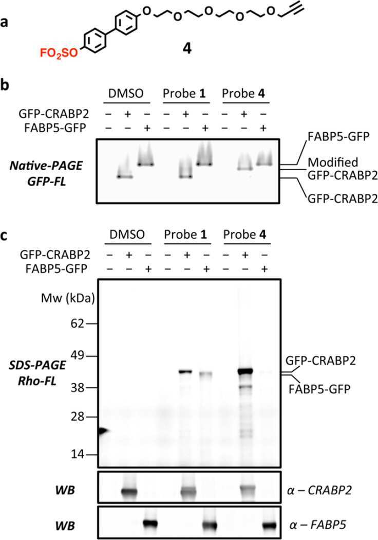
Submitted references Arylfluorosulfates Inactivate Intracellular Lipid Binding Protein(s) through Chemoselective SuFEx Reaction with a Binding Site Tyr Residue.
Integrative proteomic profiling of ovarian cancer cell lines reveals precursor cell associated proteins and functional status.
Chen W, Dong J, Plate L, Mortenson DE, Brighty GJ, Li S, Liu Y, Galmozzi A, Lee PS, Hulce JJ, Cravatt BF, Saez E, Powers ET, Wilson IA, Sharpless KB, Kelly JW
Journal of the American Chemical Society 2016 Jun 15;138(23):7353-64
Journal of the American Chemical Society 2016 Jun 15;138(23):7353-64
Integrative proteomic profiling of ovarian cancer cell lines reveals precursor cell associated proteins and functional status.
Coscia F, Watters KM, Curtis M, Eckert MA, Chiang CY, Tyanova S, Montag A, Lastra RR, Lengyel E, Mann M
Nature communications 2016 Aug 26;7:12645
Nature communications 2016 Aug 26;7:12645
No comments: Submit comment
Supportive validation
- Submitted by
- Invitrogen Antibodies (provider)
- Main image

- Experimental details
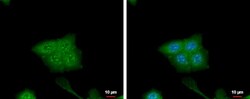
- CRABP2 Polyclonal Antibody detects CRABP2 protein at cytoplasm and nucleus by immunofluorescent analysis. Sample: HCT116 cells were fixed in 4% paraformaldehyde at RT for 15 min. Green: CRABP2 protein stained by CRABP2 Polyclonal Antibody (Product # PA5-27451) diluted at 1:500. Blue: Hoechst 33342 staining.

- Submitted by
- Invitrogen Antibodies (provider)
- Main image
- Experimental details
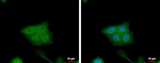
- CRABP2 Polyclonal Antibody detects CRABP2 protein at cytoplasm and nucleus by immunofluorescent analysis. Sample: HCT116 cells were fixed in 4% paraformaldehyde at RT for 15 min. Green: CRABP2 protein stained by CRABP2 Polyclonal Antibody (Product # PA5-27451) diluted at 1:500. Blue: Hoechst 33342 staining.
Supportive validation
- Submitted by
- Invitrogen Antibodies (provider)
- Main image
- Experimental details
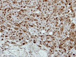
- Immunohistochemical analysis of paraffin-embedded MDAMB468 xenograft, using CRABP2 (Product # PA5-27451) antibody at 1:500 dilution. Antigen Retrieval: EDTA based buffer, pH 8.0, 15 min.

- Submitted by
- Invitrogen Antibodies (provider)
- Main image
- Experimental details
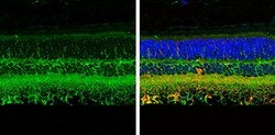
- Immunohistochemistry (Paraffin) analysis of CRABP2 was performed in paraffin-Embedded adult mouse retina tissue using Green: NF-H Polyclonal Antibody (Product # PA5-34759) at a dilution of 1:250. Red: beta Tubulin 3/ TUJ1, stained by beta Tubulin 3/ TUJ1 antibody diluted at 1:500. Blue: Fluoroshield with DAPI.

Supportive validation
- Submitted by
- Invitrogen Antibodies (provider)
- Main image
- Experimental details
- NULL
- Submitted by
- Invitrogen Antibodies (provider)
- Main image
- Experimental details
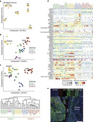
- Figure 4 Integrative analysis of HGSOC tissue and cell line proteomes. ( a ) Proteomic clustering of eight HGSOC tissue specimens. PCA was performed on the ovarian tumour specimens ( n =8) based on the proteomic profiles to evaluate inter-patient heterogeneity and intra-patient homogeneity. Component 1 and component 2 account for 51.5% of the total data variation. ( b ) PCA clustering of cell lines ( n =30), FTEC ( n =3) and HGSOC tissue proteomes ( n =8). Groups I (green), II (blue) and III (red) are as defined in Fig. 3 . The juxtaposition of the IOSE cell lines, red circles outlined in black, relative to the rest of the group III cell lines is indicated. Component 1 and component 2 account for 28.1% of the total data variation. ( c ) PCA segregation and clustering of all samples based on the 67-protein signature. The juxtaposition of the IOSE cell lines, red circles outlined in black, relative to the rest of the group III cell lines is indicated. Component 1 and component 2 account for 54.8% of the total data variation. The dendrogram below the PCA summarizes the hierarchical clustering analysis of all samples based on the 67-protein signature. Two main clusters were obtained based on this signature (detailed in Supplementary Fig. 3c ). ( d ) Relative levels of the 67 proteins used for PCA clustering in c . Relative protein levels (MaxLFQ intensities, log2) are depicted by circle size. Colours indicate protein sequence coverage per sample. MSLN is depicted twice, represent
