 Explore
 Validate
 Learn
Learn Western blot
 Immunohistochemistry
Antibody data
- Antibody Data
- Antigen structure
- References [1]
- Comments [0]
- Validations
- Immunohistochemistry [1]
Submit
Validation data
Reference
Comment
Report error
- Product number
- HPA068384 - Provider product page

- Provider
- Atlas Antibodies
- Proper citation
- Atlas Antibodies Cat#HPA068384, RRID:AB_2685979
- Product name
- Anti-CD5L
- Antibody type
- Polyclonal
- Description
- Polyclonal Antibody against Human CD5L, Gene description: CD5 molecule-like, Alternative Gene Names: API6, Spalpha, Validated applications: WB, IHC, Uniprot ID: O43866, Storage: Store at +4°C for short term storage. Long time storage is recommended at -20°C.
- Reactivity
- Human
- Host
- Rabbit
- Conjugate
- Unconjugated
- Isotype
- IgG
- Vial size
- 100 µl
- Concentration
- 0.1 mg/ml
- Storage
- Store at +4°C for short term storage. Long time storage is recommended at -20°C.
- Handling
- The antibody solution should be gently mixed before use.
Submitted references Macrophage CD5L is a target for cancer immunotherapy
Sanchez-Moral L, Paul T, Martori C, Font-Díaz J, Sanjurjo L, Aran G, Téllez É, Blanco J, Carrillo J, Ito M, Tuttolomondo M, Ditzel H, Fumagalli C, Tapia G, Sidorova J, Masnou H, Fernández-Sanmartín M, Lozano J, Vilaplana C, Rodriguez-Cortés A, Armengol C, Valledor A, Kremer L, Sarrias M
eBioMedicine 2023;91
eBioMedicine 2023;91
No comments: Submit comment
Supportive validation
- Submitted by
- Atlas Antibodies (provider)
- Enhanced method
- Orthogonal validation
- Main image
- Experimental details
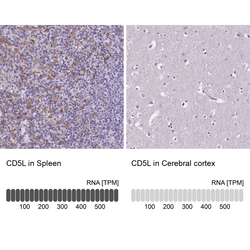
- Immunohistochemistry analysis in human spleen and cerebral cortex tissues using HPA068384 antibody. Corresponding CD5L RNA-seq data are presented for the same tissues.
- Sample type
- Human
- Protocol
- Protocol
