 Explore
 Validate
 Learn
Learn Immunoprecipitation
 Flow cytometry
Antibody data
- Antibody Data
- Antigen structure
- References [4]
- Comments [0]
- Validations
- Flow cytometry [2]
- Other assay [1]
Submit
Validation data
Reference
Comment
Report error
- Product number
- 11-0015-42 - Provider product page

- Provider
- Invitrogen Antibodies
- Product name
- CD1c Monoclonal Antibody (L161), FITC, eBioscience™
- Antibody type
- Monoclonal
- Antigen
- Other
- Description
- Description: This L161 monoclonal antibody detects CD1c (also known as BDCA-1), a glycoprotein that is noncovalently linked to beta-2 microglobulin on thymocytes and antigen presenting cells such as dendritic and Langerhans cells. This molecule is also expressed on some circulating and marginal zone B cells, as well as in lymph nodes and germinal centers. CD1c is involved in the presentation of lipid antigens such as microbial fatty acids to effector T cells during the adaptive immune response. Finally, alternative splicing gives rise to three different isoforms of CD1c (soluble, membrane, and cytoplasmic/soluble isoforms). Applications Reported: This L161 antibody has been reported for use in flow cytometric analysis, immunoprecipitation, and immunohistochemical staining. Applications Tested: This L161 antibody has been pre-titrated and tested by flow cytometric analysis on normal human peripheral blood cells. This can be used at 5 µL (0.06 µg) per test. A test is defined as the amount (µg) of antibody that will stain a cell sample in a final volume of 100 µL. Cell number should be determined empirically but can range from 10^5 to 10^8 cells/test. Excitation: 488 nm; Emission: 520 nm; Laser: Blue Laser. Filtration: 0.2 µm post-manufacturing filtered.
- Reactivity
- Human
- Host
- Mouse
- Conjugate
- Green dye
- Isotype
- IgG
- Antibody clone number
- L161
- Vial size
- 100 Tests
- Concentration
- 5 μL/Test
- Storage
- 4°C, store in dark, DO NOT FREEZE!
Submitted references Oncolytic measles virus induces tumor necrosis factor-related apoptosis-inducing ligand (TRAIL)-mediated cytotoxicity by human myeloid and plasmacytoid dendritic cells.
The Phenotypic Characterization of the Human Renal Mononuclear Phagocytes Reveal a Co-Ordinated Response to Injury.
Malaria-induced NLRP12/NLRP3-dependent caspase-1 activation mediates inflammation and hypersensitivity to bacterial superinfection.
Dendritic cells activated by IFN-γ/STAT1 express IL-31 receptor and release proinflammatory mediators upon IL-31 treatment.
Achard C, Guillerme JB, Bruni D, Boisgerault N, Combredet C, Tangy F, Jouvenet N, Grégoire M, Fonteneau JF
Oncoimmunology 2017;6(1):e1261240
Oncoimmunology 2017;6(1):e1261240
The Phenotypic Characterization of the Human Renal Mononuclear Phagocytes Reveal a Co-Ordinated Response to Injury.
Leone DA, Kozakowski N, Kornauth C, Waidacher T, Neudert B, Loeffler AG, Haitel A, Rees AJ, Kain R
PloS one 2016;11(3):e0151674
PloS one 2016;11(3):e0151674
Malaria-induced NLRP12/NLRP3-dependent caspase-1 activation mediates inflammation and hypersensitivity to bacterial superinfection.
Ataide MA, Andrade WA, Zamboni DS, Wang D, Souza Mdo C, Franklin BS, Elian S, Martins FS, Pereira D, Reed G, Fitzgerald KA, Golenbock DT, Gazzinelli RT
PLoS pathogens 2014 Jan;10(1):e1003885
PLoS pathogens 2014 Jan;10(1):e1003885
Dendritic cells activated by IFN-γ/STAT1 express IL-31 receptor and release proinflammatory mediators upon IL-31 treatment.
Horejs-Hoeck J, Schwarz H, Lamprecht S, Maier E, Hainzl S, Schmittner M, Posselt G, Stoecklinger A, Hawranek T, Duschl A
Journal of immunology (Baltimore, Md. : 1950) 2012 Jun 1;188(11):5319-26
Journal of immunology (Baltimore, Md. : 1950) 2012 Jun 1;188(11):5319-26
No comments: Submit comment
Supportive validation
- Submitted by
- Invitrogen Antibodies (provider)
- Main image

- Experimental details
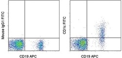
- Staining of normal human peripheral blood cells with Anti-Human CD19 APC (Product # 17-0199-42) and Mouse IgG1 K Isotype Control FITC (Product # 11-4714-42) (left) or Anti-Human CD1c (right). Total viable cells were used for this analysis.
- Conjugate
- Green dye

- Submitted by
- Invitrogen Antibodies (provider)
- Main image
- Experimental details
- Staining of normal human peripheral blood cells with Anti-Human CD19 APC (Product # 17-0199-42) and Mouse IgG1 K Isotype Control FITC (Product # 11-4714-42) (left) or Anti-Human CD1c (right). Total viable cells were used for this analysis.

Supportive validation
- Submitted by
- Invitrogen Antibodies (provider)
- Main image
- Experimental details
- NULL
- Conjugate
- Green dye
