Explore
 Validate
 Learn
Learn Western blot
 Immunoprecipitation
Antibody data
- Antibody Data
- Antigen structure
- References [0]
- Comments [0]
- Validations
- Western blot [3]
Submit
Validation data
Reference
Comment
Report error
- Product number
- MA5-14942 - Provider product page

- Provider
- Invitrogen Antibodies
- Product name
- LKB1 Monoclonal Antibody (B.593.0)
- Antibody type
- Monoclonal
- Antigen
- Synthetic peptide
- Description
- It is not recommended to aliquot this antibody.
- Reactivity
- Human
- Host
- Rabbit
- Isotype
- IgG
- Antibody clone number
- B.593.0
- Vial size
- 100 µL
- Storage
- -20°C
No comments: Submit comment
Supportive validation
- Submitted by
- Invitrogen Antibodies (provider)
- Main image

- Experimental details
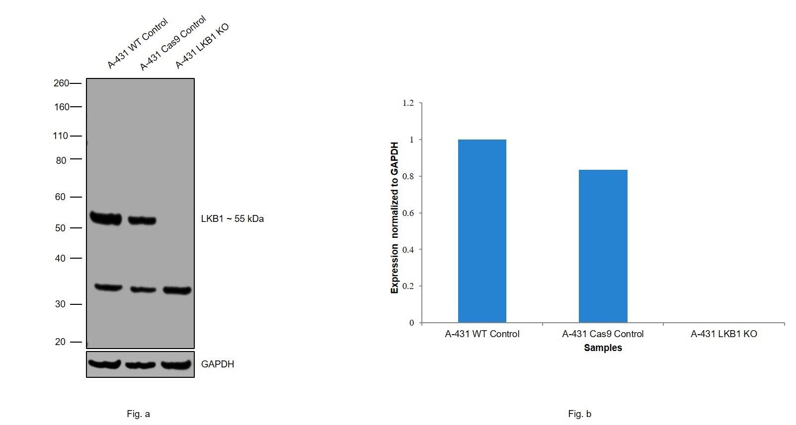
- Knockout of LKB1 was achieved by CRISPR-Cas9 genome editing using LentiArray™ Lentiviral sgRNA (Product # A32042, AssayID CRISPR870103_LV) and LentiArray Cas9 Lentivirus (Product # A32064). Western blot analysis of LKB1 was performed by loading 30 µg of A-431 wild type (Lane 1), A-431 CAS9 (Lane 2), A-431 LKB1 KO (Lane 3) whole cell extracts. The blot was probed with Anti-LKB1 Monoclonal Antibody (B.593.0)(Product # MA5-14942) using 1:1000 dilution and Goat anti-Rabbit IgG (H+L), Superclonal™ Recombinant Secondary Antibody, HRP (Product # A27036). Loss of signal upon CRISPR mediated knockout (KO) using the LentiArray™ CRISPR product line confirms that antibody is specific to LKB1. An uncharacterized band was observed at ~32kDa.
- Submitted by
- Invitrogen Antibodies (provider)
- Main image
- Experimental details
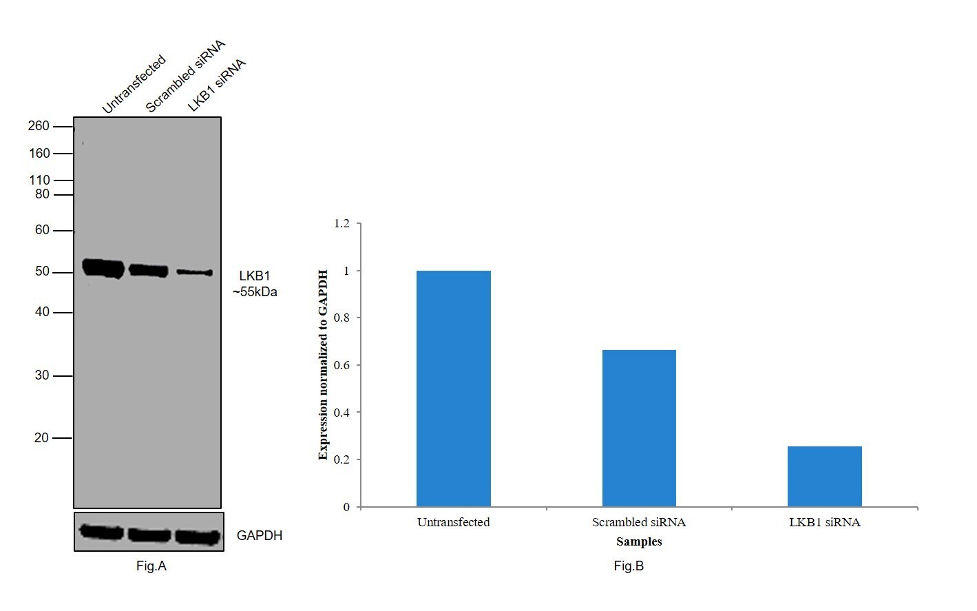
- Knockdown of LKB1 was achieved by transfecting A-431 with LKB1 specific siRNAs (Silencer® select Product # s532020, s501375). Western blot analysis (Fig. a) was performed using whole cell extracts from the LKB1 knockdown cells (Lane 3), non-specific scrambled siRNA transfected cells (Lane 2) and untransfected cells (Lane 1). The blot was probed with LKB1 Monoclonal Antibody (B.593.0) (Product # MA5-14942, 1:1000 dilution) and Goat anti-Rabbit IgG (H+L) Superclonal™ Recombinant Secondary Antibody, HRP (Product # A27036, 1:4000 dilution). Densitometric analysis of this western blot is shown in histogram (Fig. b). Decrease in signal upon siRNA mediated knock down confirms that antibody is specific to LKB1.
- Submitted by
- Invitrogen Antibodies (provider)
- Main image
- Experimental details
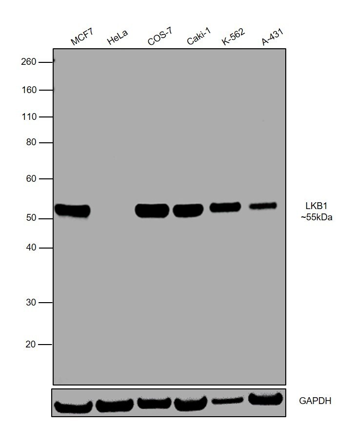
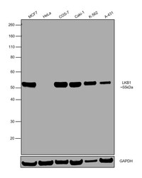
- Western blot was performed using Anti-LKB1 Monoclonal Antibody (B.593.0) (Product # MA5-14942) and a 55kDa band corresponding to LKB1 was observed in all the tested cell models. Modified whole cell lysate (1% SDS) (30ug lysate) of MCF7 (Lane 1), HeLa (Lane 2), COS-7 (Lane 3), Caki-1 (Lane 4), K-562 (Lane 5), and A-431 (Lane 6) were electrophoresed using Novex® NuPAGE® 4-12 % Bis-Tris gel (Product # NP0322BOX). Resolved proteins were then transferred onto a nitrocellulose membrane (Product # IB23001) by iBlot® 2 Dry Blotting System (Product # IB21001). The blot was probed with the primary antibody (1:1000 dilution) and detected by chemiluminescence with Goat anti-Rabbit IgG (H+L), Superclonal™ Recombinant Secondary Antibody, HRP conjugate (Product # A27036, 1:4000 dilution) using the iBright FL 1000 (Product # A32752). Chemiluminescent detection was performed using Novex® ECL Chemiluminescent Substrate Reagent Kit (Product # WP20005).