Explore
 Validate
 Learn
Learn Western blot
 ELISA
ELISA Immunocytochemistry
Antibody data
- Antibody Data
- Antigen structure
- References [1]
- Comments [0]
- Validations
- Immunocytochemistry [4]
- Immunohistochemistry [1]
- Other assay [1]
Submit
Validation data
Reference
Comment
Report error
- Product number
- PA5-96062 - Provider product page

- Provider
- Invitrogen Antibodies
- Product name
- LKB1 Polyclonal Antibody
- Antibody type
- Polyclonal
- Antigen
- Recombinant full-length protein
- Description
- Immunogen sequence: CGMQEMLDSV PEKRFPVCQA HGYFCQLIDG LEYLHSQGIV HKDIKPGNLL LTTGGTLKIS DLGVAEALHP FAADDTCRTS QGSPAFQPPE IANGLDTFSG FKVDIWSAGV TLYNITTGLY PFEGDNIYKL FENIGKGSYA IPGDCGPPLS DLLKGMLEYE PAKRFSIRQI RQHSWFRKKH PPAEAPVPIP PSPDTKDRWR SMTVVPYLED LHGADEDEDL FDIEDDIIYT QDFTVPGQVP EEEASHNGQR RGLPKAVCMN GTEAAQLSTK SRAEGRAPNP ARKACSASSK IRRLSACKQQ; Positive Samples: U-87MG, SKOV3, Mouse heart; Cellular Location: Cytoplasm, Membrane, Mitochondrion, Nucleus
- Reactivity
- Human, Mouse, Rat
- Host
- Rabbit
- Isotype
- IgG
- Vial size
- 100 μL
- Concentration
- 0.75 mg/mL
- Storage
- -20°C, Avoid Freeze/Thaw Cycles
Submitted references Low-glucose-sensitive TRPC6 dysfunction drives hypoglycemia-induced cognitive impairment in diabetes.
He C, Gao P, Cui Y, Li Q, Li Y, Lu Z, Ma H, Zhao Y, Li L, Sun F, Chen X, Jia H, Liu D, Yang G, Zheng H, Zhu Z
Clinical and translational medicine 2020 Oct;10(6):e205
Clinical and translational medicine 2020 Oct;10(6):e205
No comments: Submit comment
Supportive validation
- Submitted by
- Invitrogen Antibodies (provider)
- Main image
- Experimental details
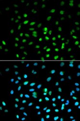
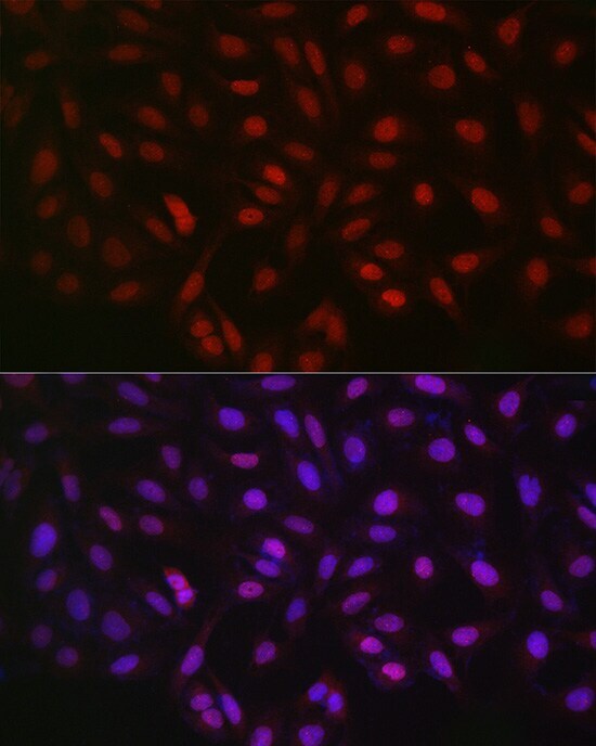
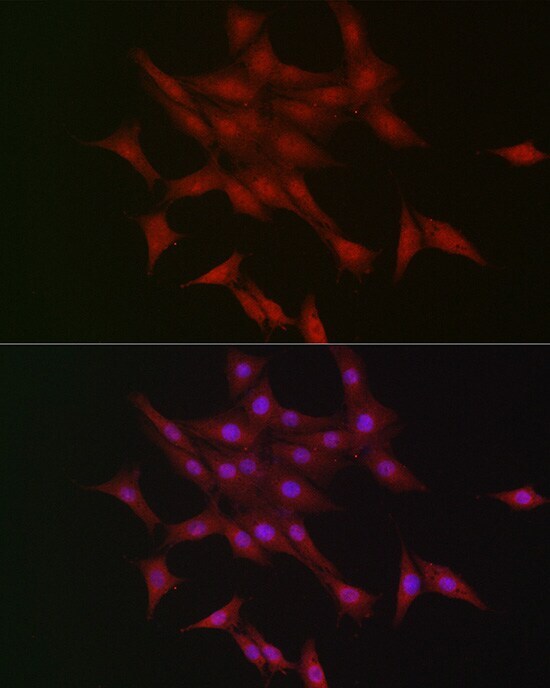
- Immunocytochemistry-Immunofluorescence analysis of LKB1 was performed in MCF-7 cells using LKB1 Polyclonal Antibody (Product # PA5-96062). Blue: DAPI for nuclear staining.
- Submitted by
- Invitrogen Antibodies (provider)
- Main image
- Experimental details
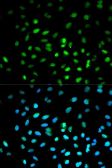
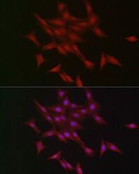
- Immunofluorescence analysis of LKB1 in U2OS cells. Samples were incubated with LKB1 Polyclonal antibody (Product # PA5-96062) using a dilution of 1:50 (40x lens). Blue: DAPI for nuclear staining.
- Submitted by
- Invitrogen Antibodies (provider)
- Main image
- Experimental details
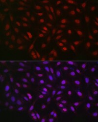
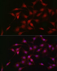
- Immunofluorescence analysis of LKB1 in NIH/3T3 cells. Samples were incubated with LKB1 Polyclonal antibody (Product # PA5-96062) using a dilution of 1:50 (40x lens). Blue: DAPI for nuclear staining.
- Submitted by
- Invitrogen Antibodies (provider)
- Main image
- Experimental details
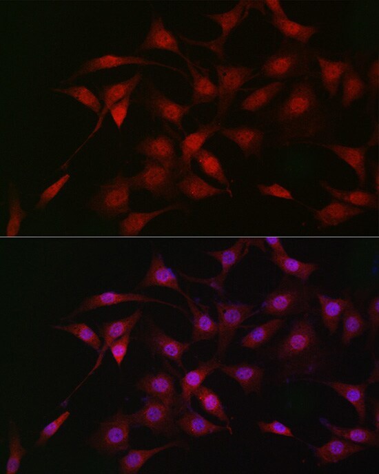
- Immunofluorescence analysis of LKB1 in PC-12 cells. Samples were incubated with LKB1 Polyclonal antibody (Product # PA5-96062) using a dilution of 1:50 (40x lens). Blue: DAPI for nuclear staining.
Supportive validation
- Submitted by
- Invitrogen Antibodies (provider)
- Main image

- Experimental details
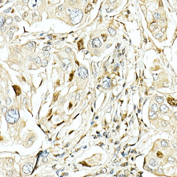
- Immunohistochemistry analysis of LKB1 in paraffin-embedded human lung cancer. Samples were incubated with LKB1 Polyclonal antibody (Product # PA5-96062) using a dilution of 1:50 (40x lens). Perform high pressure antigen retrieval with 10 mM citrate buffer pH 6.0 before commencing with IHC staining protocol.
Supportive validation
- Submitted by
- Invitrogen Antibodies (provider)
- Main image
- Experimental details
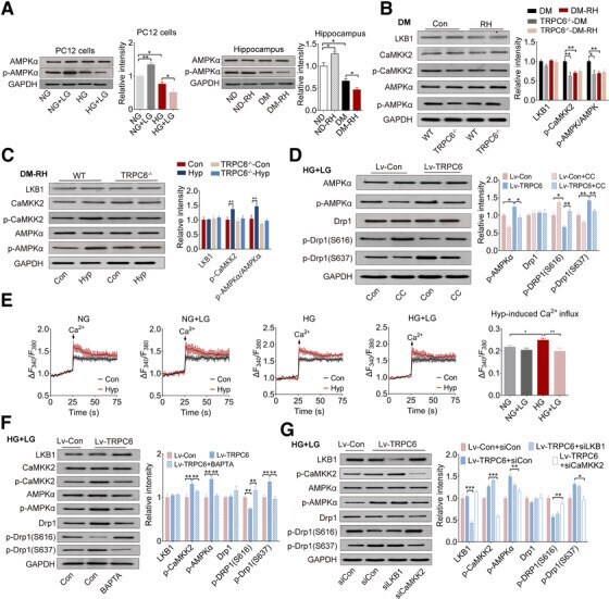
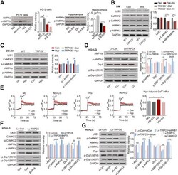
- FIGURE 6 Inhibition of AMPK by TRPC6/ CaMKK2 pathway disturbs phosphorylation of Drp1. (A) Representative Western blots of AMPKalpha and p-AMPKalpha in PC12 cells or mouse hippocampus. The quantitative data are showed on the right (n = 3 for PC12 cells, n = 4 tissues from four mice). (B) Representative Western blots of LKB1, CaMKK2, p-CaMKK2, AMPKalpha, and p-AMPKalpha in the hippocampus of DM mice with or without RH. The quantitative data are shown on the right (n = 3 tissues from three mice). TRPC6 -/- , TRPC6 global knockout mice. (C) Representative Western blots of LKB1, CaMKK2, p-CaMKK2, AMPKalpha, and p-AMPKalpha in the hippocampus of hyperforin or vehicle-treated diabetic WT or TRPC6 -/- -mice with RH. The quantitative data are showed on the right (n = 3). Hyp, hyperforin. (D) The protein expression level of AMPKalpha, p-AMPKalpha, Drp1, p-Drp1, (S616), and p-Drp1 (S637) in PC12 cells treated with CC. The statistical results are showed on the right (n = 3). CC, AMPK inhibitor compound C (10 uM); Lv-TRPC6, TRPC6 overexpression with lentivirus; Lv-Con, the vehicle lentivirus. (E) The hyperforin (10 uM) evoked intracellular Ca 2+ influx in PC12 cells cultured. The quantitative data are showed on the right (n = 3). TRPC6-mediated Ca 2+ influx was determined by the subtraction of hyperforin- and Ca 2+ -induced Ca 2+ influx to Ca 2+ -induced influx. Ca 2+ (1 mM) and/or hypforin (10 uM) was added at 25S. Ca 2+ , Cacl 2 (1 mM); Hyp, hyperforim (10 uM). (F) The protein levels o