Explore
 Validate
 Learn
Learn Western blot
 Immunocytochemistry
Antibody data
- Antibody Data
- Antigen structure
- References [0]
- Comments [0]
- Validations
- Immunocytochemistry [1]
- Immunoprecipitation [1]
Submit
Validation data
Reference
Comment
Report error
- Product number
- PA5-115222 - Provider product page

- Provider
- Invitrogen Antibodies
- Product name
- FCER1G Polyclonal Antibody
- Antibody type
- Polyclonal
- Antigen
- Synthetic peptide
- Description
- Antibody detects endogenous levels of total FCER1G.
- Reactivity
- Human, Mouse, Rat
- Host
- Rabbit
- Isotype
- IgG
- Vial size
- 100 μL
- Concentration
- 1 mg/mL
- Storage
- -20°C
No comments: Submit comment
Supportive validation
- Submitted by
- Invitrogen Antibodies (provider)
- Main image
- Experimental details
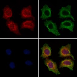
- Immunocytochemistry analysis of FCER1G in HepG2 cells. Samples were treated with PFA, permeabilized in 0.1% Triton X-100, blocked in 10% serum (45 min at 25°C), and incubated with polyclonal antibody (Product # PA5-115222) at a dilution of 1:200 (1 hr at 37°C). Secondary staining was applied with mouse anti-beta tubulin; AlexaFluor 594 conjugated goat anti-rabbit IgG (Red); AlexaFluor 88 conjugated goat anti-mouse IgG (Green) and DAPI (blue) using a dilution of 1:200 (1 hr at 37°C).
Supportive validation
- Submitted by
- Invitrogen Antibodies (provider)
- Main image
- Experimental details
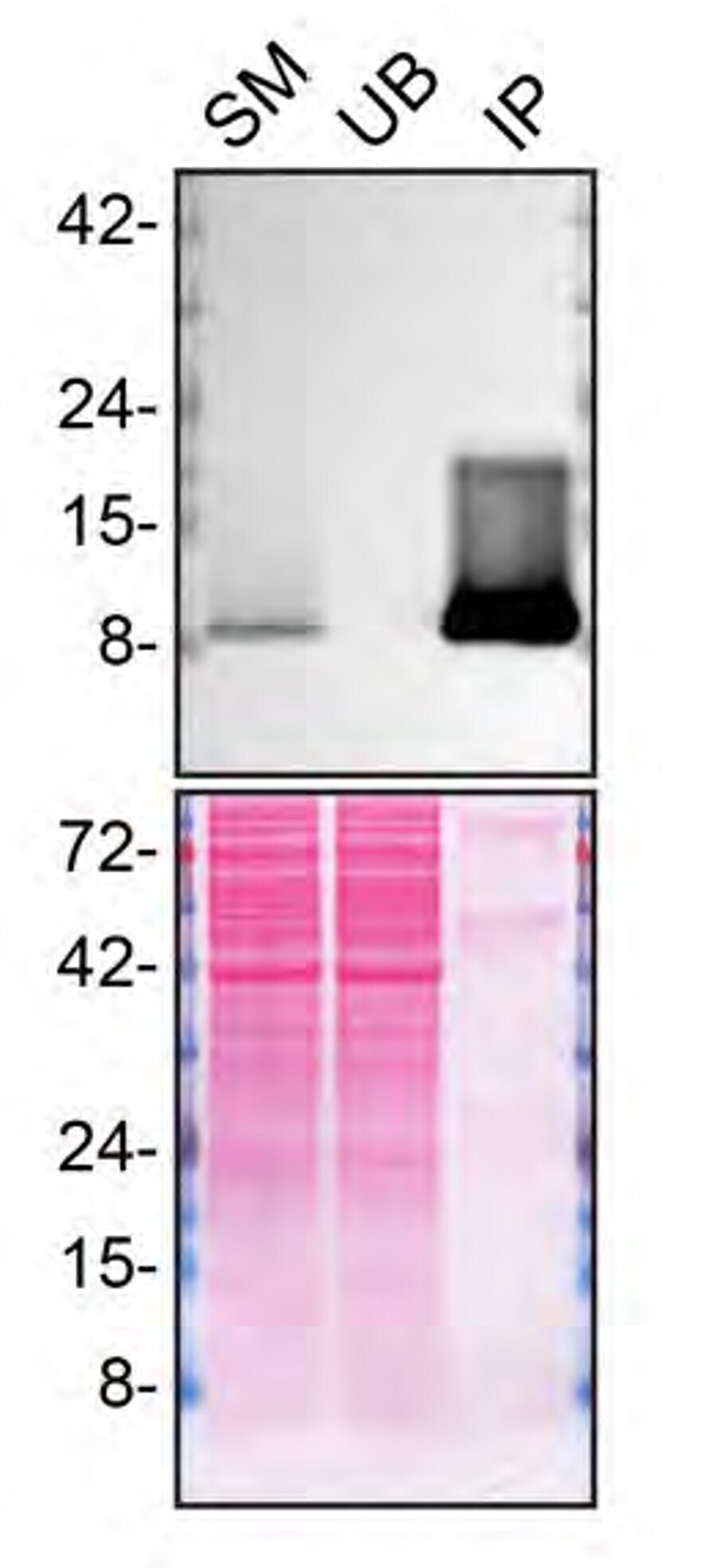
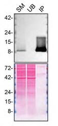
- Immunoprecipitation of FCER1G was performed on PMA-treated THP-1 WT cell lysate. Antibody-bead conjugate was prepared by adding 2 µg of FCER1G Polyclonal Antibody (Product # PA5-115222) and 30 µL of Dynabeads™ Protein A (Product # 10002D) to 500 μl of Pierce™ IP Lysis Buffer (Product # 87788). The mixture was rocked for ~1 hour at 4 degree celcius followed by two washes to remove unbound antibodies. Cells were lysed in Pierce™ IP Lysis Buffer (Product # 87788) supplemented with protease inhibitor. The lysate was rocked for 30 min at 4 degrees Celsius and spun at 110,000xg for 15 min at 4 degrees Celsius. 600 µg of lysate was incubated with the antibody-bead conjugate for ~1 hours at 4 degrees Celsius. Following centrifugation and multiple washes, 6% starting material (SM), 6% unbound fraction (UB) and immunoprecipitated fraction (IP) were processed for immunoblot using a different antibody. Ponceau stained transfer of blot is shown (below immunoblot). Data courtesy of YCharOS Inc., an open science company with the mission of characterizing commercially available antibodies using knockout validation.