Explore
 Validate
 Learn
Learn Western blot
 ELISA
ELISAAntibody data
- Antibody Data
- Antigen structure
- References [0]
- Comments [0]
- Validations
- Western blot [1]
- Immunocytochemistry [2]
Submit
Validation data
Reference
Comment
Report error
- Product number
- PA1-26900 - Provider product page

- Provider
- Invitrogen Antibodies
- Product name
- ApoA2 Polyclonal Antibody
- Antibody type
- Polyclonal
- Antigen
- Other
- Description
- PA1-26900 detects Apolipoprotein A II from human samples.
- Reactivity
- Human
- Host
- Goat
- Isotype
- IgG
- Vial size
- 100 µg
- Concentration
- 1 mg/mL
- Storage
- Store at 4°C short term. For long term storage, store at -20°C, avoiding freeze/thaw cycles.
No comments: Submit comment
Supportive validation
- Submitted by
- Invitrogen Antibodies (provider)
- Main image
- Experimental details
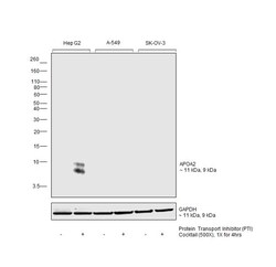
- Western blot was performed using Anti-ApoA2 Polyclonal Antibody (Product # PA1-26900) and a 11kDa band corresponding to Apolipoprotein A-II was observed along with a band at 9 kDa which is one of the isoform observed across cell lines tested. Whole cell extracts (40 µg lysate) of Hep G2 (Lane 1), Hep G2 treated with PTI (1X for 4hrs) (Lane 2), A549 (Lane 3), A549 treated with PTI (1X for 4hrs) (Lane 4), SK-O-V3 (Lane 5), SK-O-V3 treated with PTI (1X for 4hrs) (Lane 6) were electrophoresed using Novex™ 16% Tricine Protein Gel (Product # EC6695BOX). Resolved proteins were then transferred onto a nitrocellulose membrane (Product # IB23001) by iBlot® 2 Dry Blotting System (Product # IB21001). The blot was probed with the primary antibody (1:5000 dilution) and detected by chemiluminescence with Rabbit anti-Goat IgG (H+L) Superclonal™ Recombinant Secondary Antibody, HRP (Product # A27014,1:20000 dilution) using the iBright FL 1000 (Product # A32752). Chemiluminescent detection was performed using SuperSignal™ West Pico PLUS Chemiluminescent Substrate (Product # 34580).

Supportive validation
- Submitted by
- Invitrogen Antibodies (provider)
- Main image
- Experimental details
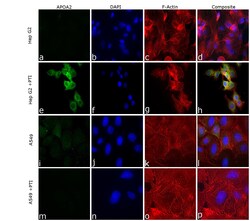
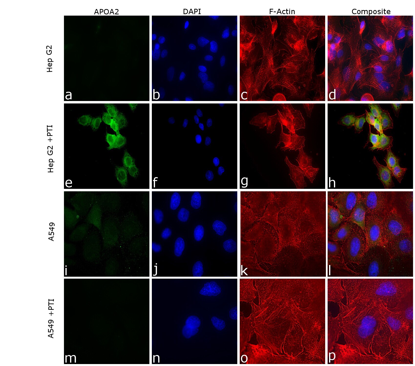
- Immunofluorescence analysis of Apolipoprotein A-II was performed using 70% confluent log phase Hep G2 cells treated with PTI (1X for 4hrs) and A-549 cells treated with PTI (1X for 4hrs). The cells were fixed with 4% paraformaldehyde for 10 minutes, permeabilized with 0.1% Triton™ X-100 for 15 minutes, and blocked with 2% BSA for 45 minutes at room temperature. The cells were labeled with ApoA2 Polyclonal Antibody (Product # PA1-26900) at 1:100 dilution in 0.1% BSA, incubated at 4 degree celsius overnight and then labeled with Rabbit anti-Goat IgG (H+L) Cross-Adsorbed Secondary Antibody, Alexa Fluor 488 (Product # A11078), (1:2000 dilution), for 45 minutes at room temperature (Panel a, e ,i, m: Green). Nuclei (Panel b, f, j, n: Blue) were stained with ProLong™ Diamond Antifade Mountant with DAPI (Product # P36962). F-actin (Panel c, g, k, o: Red) was stained with Rhodamine Phalloidin (Product # R415, 1:300 dilution). Panel h represents the merged image showing cytoplasmic localization in Hep G2 cells treated with PTI. Panel d and l represents untreated cells of Hep G2 and A549 cells respectively, Panel P representes A549 cells treated with PTI. The images were captured at 60X magnification in EVOS™ M7000 Imaging System (Product # AMF7000) and externally deconvoluted (D.Sage et al. / Methods 115 (2017) 28-41).
- Submitted by
- Invitrogen Antibodies (provider)
- Main image

- Experimental details
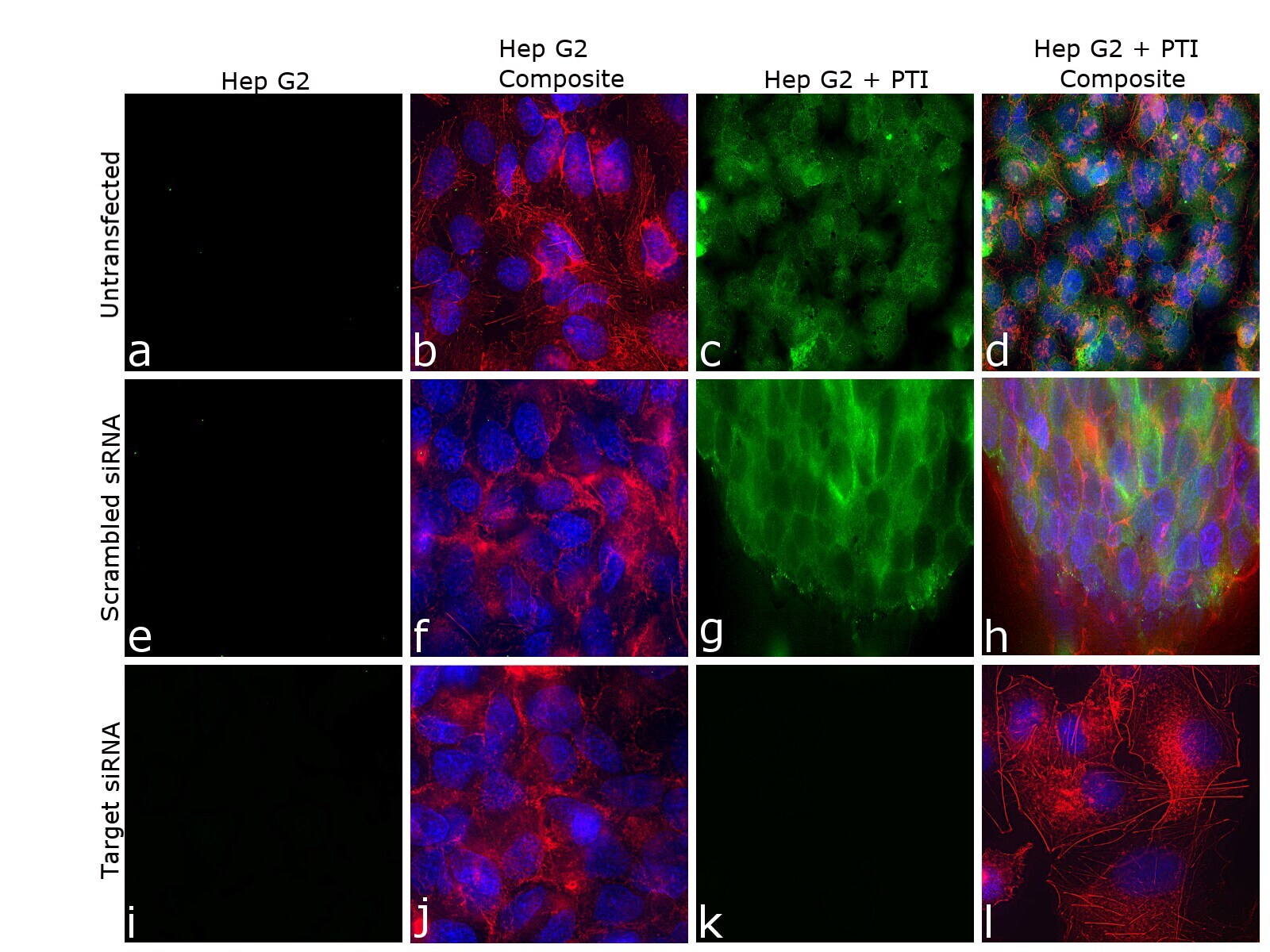
- Knockdown of Apolipoprotein A-II was achieved by transfecting Hep G2 cells with Apolipoprotein A-II specific siRNA (Silencer® select Product # s1470, s1471).Immunofluorescence analysis was performed on untransfected Hep G2 cells (panel a,b), untransfected Hep G2 cells treated with PTI (1X for 4hrs) (panel c, d), non-specific scrambled siRNA transfected Hep G2 cells (panels e,f), non-specific scrambled siRNA transfected Hep G2 cells treated with PTI (1X for 4hrs) (panels g,h), Hep G2 cells transfected with Apolipoprotein A-II specific siRNA (panel i,j), and Hep G2 cells transfected with Apolipoprotein A-II specific siRNA and then treated with PTI (1X for 4hrs) (panels k,l) (Green) Cells were fixed, permeabilized, and labelled with ApoA2 Polyclonal Antibody (Product # PA1-26900, 5 µg/mL) followed by Rabbit anti-Goat IgG (H+L) Cross-Adsorbed Secondary Antibody, Alexa Fluor 488 (Product # A11078), (1:2000 dilution). Nuclei (blue) were stained using ProLong™ Diamond Antifade Mountant with DAPI (Product # P36962), and Rhodamine Phalloidin (Product # R415, 1:300 dilution) was used for cytoskeletal F-actin (Green) staining. Reduction in specific signal was observed upon siRNA mediated knockdown (panel k ,l) confirming specificity of the antibody to Apolipoprotein A-II .The images were captured at 60X magnification in EVOS™ M7000 Imaging System (Product # AMF7000) and externally deconvoluted (D.Sage et al. / Methods 115 (2017) 28-41).