 Explore
 Validate
 Learn
Learn Western blot
Antibody data
- Antibody Data
- Antigen structure
- References [0]
- Comments [0]
- Validations
- Western blot [1]
- Immunocytochemistry [3]
Submit
Validation data
Reference
Comment
Report error
- Product number
- 701236 - Provider product page

- Provider
- Invitrogen Antibodies
- Product name
- ApoA2 Recombinant Rabbit Monoclonal Antibody (43H22L4)
- Antibody type
- Monoclonal
- Antigen
- Synthetic peptide
- Reactivity
- Human, Rat
- Host
- Rabbit
- Isotype
- IgG
- Antibody clone number
- 43H22L4
- Vial size
- 100 µg
- Concentration
- 0.5 mg/mL
- Storage
- Store at 4°C short term. For long term storage, store at -20°C, avoiding freeze/thaw cycles.
No comments: Submit comment
Supportive validation
- Submitted by
- Invitrogen Antibodies (provider)
- Main image
- Experimental details
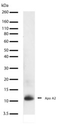
- Western blot analysis of ApoA2 in rat liver lysate using an ApoA2 recombinant rabbit monoclonal antibody (Product # 701236) at a dilution of 2 µg/mL. Samples were detected using chemiluminescence (ECL). Results show a band at ~12kDa.

Supportive validation
- Submitted by
- Invitrogen Antibodies (provider)
- Main image
- Experimental details
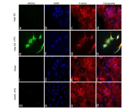
- Immunofluorescence analysis of Apolipoprotein A-II was performed using 70% confluent log phase Hep G2 cells. The cells were fixed with 4% paraformaldehyde for 10 minutes, permeabilized with 0.1% Triton™ X-100 for 15 minutes, and blocked with 2% BSA for 45 minutes at room temperature. The cells were labeled with ApoA2 Recombinant Rabbit Monoclonal Antibody (43H22L4) (Product # 701236) at 5 µg/mL in 0.1% BSA, incubated at 4 degree celsius overnight and then labeled with Donkey anti-Rabbit IgG (H+L) Highly Cross-Adsorbed Secondary Antibody, Alexa Fluor Plus 488 (Product # A32790), (1:2000 dilution), for 45 minutes at room temperature(Panel a, e ,i, m: Green). Nuclei (Panel b, f, j, n: Blue) were stained with ProLong™ Diamond Antifade Mountant with DAPI (Product # P36962). F-actin (Panel c, g, k, o: Red) was stained with Rhodamine Phalloidin (Product # R415, 1:300 dilution). Panel h represents the merged image showing cytoplasmic localization in Hep G2 cells treated with PTI. Panel d and l represents untreated cells of Hep G2 and A549 cells respectively, Panel P representes A549 cells treated with PTI. The images were captured at 60X magnification in EVOS™ M7000 Imaging System (Product # AMF7000) and externally deconvoluted (D.Sage et al. / Methods 115 (2017) 28-41).

- Submitted by
- Invitrogen Antibodies (provider)
- Main image

- Experimental details
- Knockdown of Apolipoprotein A-II was achieved by transfecting Hep G2 cells with Apolipoprotein A-II specific siRNA (Silencer® select Product # s1470, s1471). Immunofluorescence analysis was performed on untransfected Hep G2 cells (panel a,b), untransfected Hep G2 cells treated with PTI (1X for 4hrs) (panel c, d), non-specific scrambled siRNA transfected Hep G2 cells (panels e,f), non-specific scrambled siRNA transfected Hep G2 cells treated with PTI (1X for 4hrs) (panels g,h), Hep G2 cells transfected with Apolipoprotein A-II specific siRNA (panel i,j), and Hep G2 cells transfected with Apolipoprotein A-II specific siRNA and then treated with PTI (1X for 4hrs) (panels k,l) (Green) Cells were fixed, permeabilized, and labelled with ApoA2 Recombinant Rabbit Monoclonal Antibody (43H22L4) (Product # 701236, 5 µg/mL) followed by Donkey anti-Rabbit IgG (H+L) Highly Cross-Adsorbed Secondary Antibody, Alexa Fluor Plus 488 (Product # A32790), (1:2000 dilution). Nuclei (blue) were stained using ProLong™ Diamond Antifade Mountant with DAPI (Product # P36962), and Rhodamine Phalloidin (Product # R415, 1:300 dilution) was used for cytoskeletal F-actin (Green) staining. Reduction in specific signal was observed upon siRNA mediated knockdown (panel k ,l) confirming specificity of the antibody to Apolipoprotein A-II .The images were captured at 60X magnification in EVOS™ M7000 Imaging System (Product # AMF7000) and externally deconvoluted (D.Sage et al. / Methods 115 (2017) 28-41).

- Submitted by
- Invitrogen Antibodies (provider)
- Main image
- Experimental details
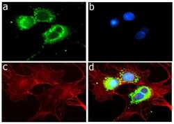
- Immunofluorescent analysis of ApoA2 in HepG2 cells using an ApoA2 recombinant rabbit monoclonal antibody (Product # 701236) followed by detection using an Alexa Fluor 488-conjugated goat anti-rabbit secondary antibody (green) (Image A). Nuclei were stained using DAPI (Image B) and actin stained with Alexa Fluor 594 phalloidin (red) (image C). Image D is a composite image showing cytoplasmic localization of Apo A2.
